
Upper Limb Venous Drainage Anatomy | Cephalic & Basilic Veins
Learn about the superficial and deep venous drainage of the upper limb, including the cephalic, basilic, and median cubital veins for clinical practice.
Venous Drainage of the Upper Limb
Anatomical Overview based on Gray's Anatomy & B.D. Chaurasia
Introduction to Venous Classification
The veins of the upper limb are divided into two primary groups: Superficial and Deep.
Superficial veins lie in the superficial fascia (subcutaneous tissue) and are commonly used for venipuncture.
Deep veins lie deep to the deep fascia and accompany major arteries as venae comitantes.
Both systems have valves to prevent backflow and communicate via perforating veins.
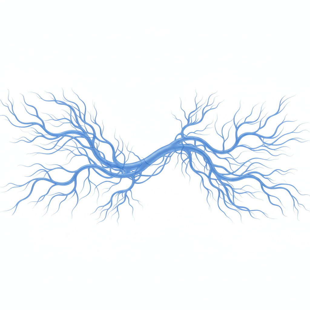
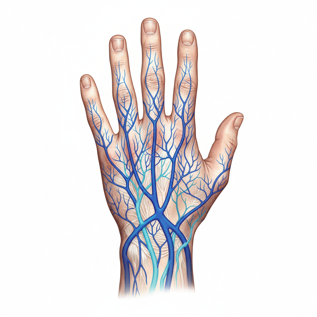
Origin: The Dorsal Venous Network
The superficial veins begin on the dorsum of the hand. The dorsal digital veins unite to form dorsal metacarpal veins, which eventually form the dorsal venous arch (or network). The lateral end of this arch gives rise to the Cephalic vein, while the medial end gives rise to the Basilic vein.
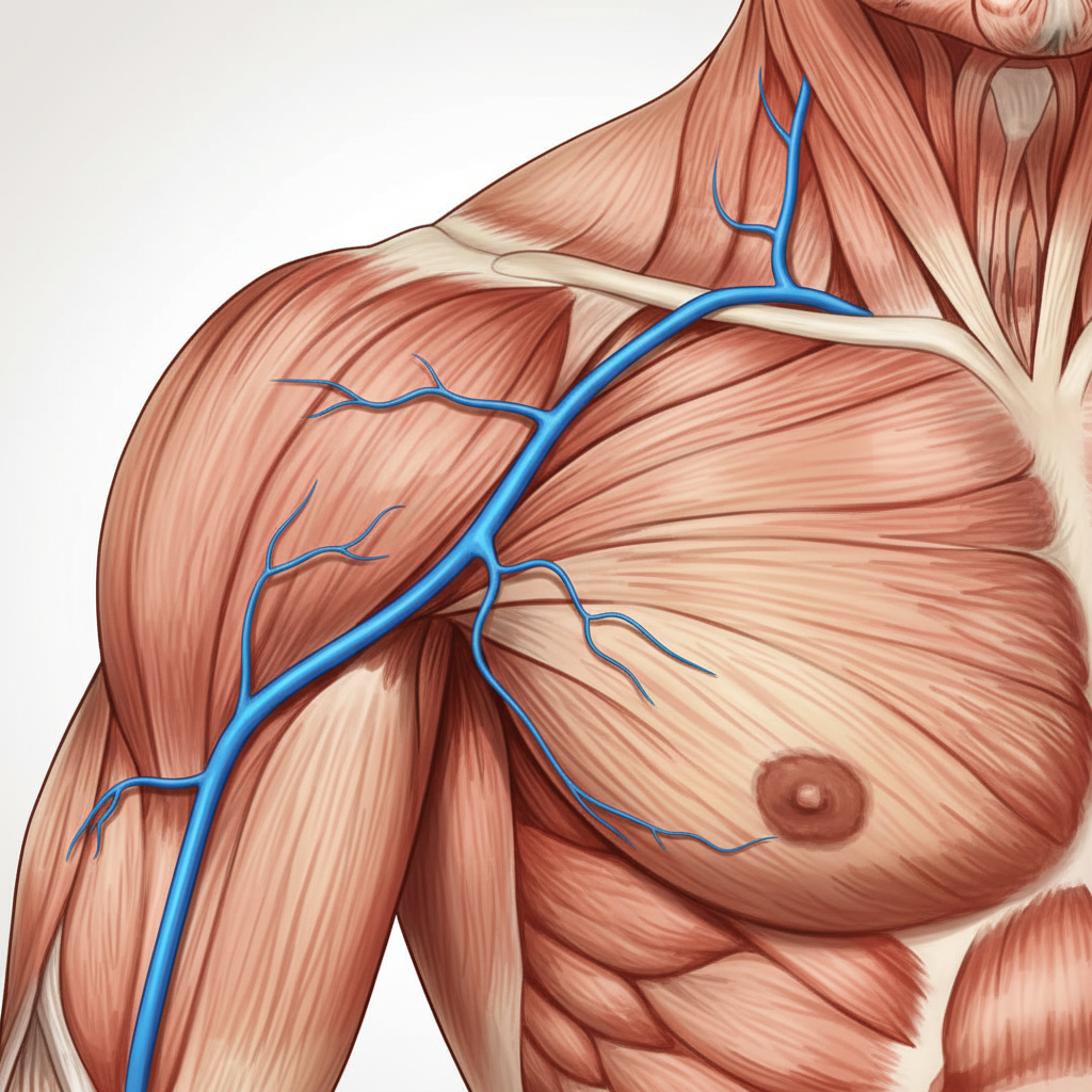
The Cephalic Vein (Pre-axial Vein)
The cephalic vein ascends along the lateral aspect of the forearm and arm. It runs in the deltopectoral groove between the deltoid and pectoralis major muscles. Finally, it pierces the clavipectoral fascia to drain into the axillary vein.
The Basilic Vein (Post-axial Vein)
Arises from the medial end of the dorsal venous arch.
Ascends along the medial aspect of the forearm and lower arm.
At the middle of the arm, it pierces the deep fascia.
Continues upwards to equal the lower border of teres major, where it becomes the axillary vein.
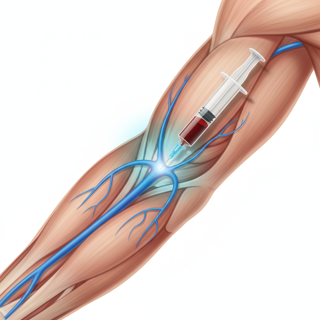
Median Cubital Vein
Located in the cubital fossa (front of the elbow), this large communicating vein connects the cephalic and basilic veins. It runs obliquely upwards and medially. Because it is fixed by the bicipital aponeurosis and is superficial, it is the vein of choice for withdrawing blood (venipuncture) and intravenous injections.
The deep veins generally accompany the arteries as venae comitantes, usually arranged as a pair of veins flanking the artery.
General Rule of Deep Drainage
The Axillary Vein
Formation: Begins at the lower border of the teres major muscle as a continuation of the basilic vein.
Course: Runs through the axilla, medial to the axillary artery.
Tributaries: Receives the cephalic vein, venae comitantes of the brachial artery, and tributaries corresponding to branches of the axillary artery.
Termination: Ends at the outer border of the first rib, becoming the Subclavian Vein.
Clinical Significance
1. Venipuncture: The median cubital vein is the most common site for drawing blood. 2. Deep Vein Thrombosis (DVT): Can occur in the axillary or subclavian veins (Paget-Schroetter syndrome). 3. Cutdown: The cephalic vein at the deltopectoral groove is a consistent site for venous cutdown procedures when other access fails.
References
Chaurasia, B. D. (2020). Human Anatomy: Upper Limb & Thorax (8th ed.). CBS Publishers & Distributors.
Standring, S. (2020). Gray's Anatomy: The Anatomical Basis of Clinical Practice (42nd ed.). Elsevier.
Moore, K. L., Dalley, A. F., & Agur, A. M. R. (2017). Clinically Oriented Anatomy.
- anatomy
- venous-drainage
- upper-limb
- cephalic-vein
- basilic-vein
- venipuncture
- medical-education
- human-biology