Heart and Blood Vessel Anatomy: Structural Overview
Explore the structural anatomy of the heart and blood vessels, including chambers, heart valves, coronary circulation, and histological vessel layers.
Total Anatomy
Structural Overview for Allied Health & Pre-Med
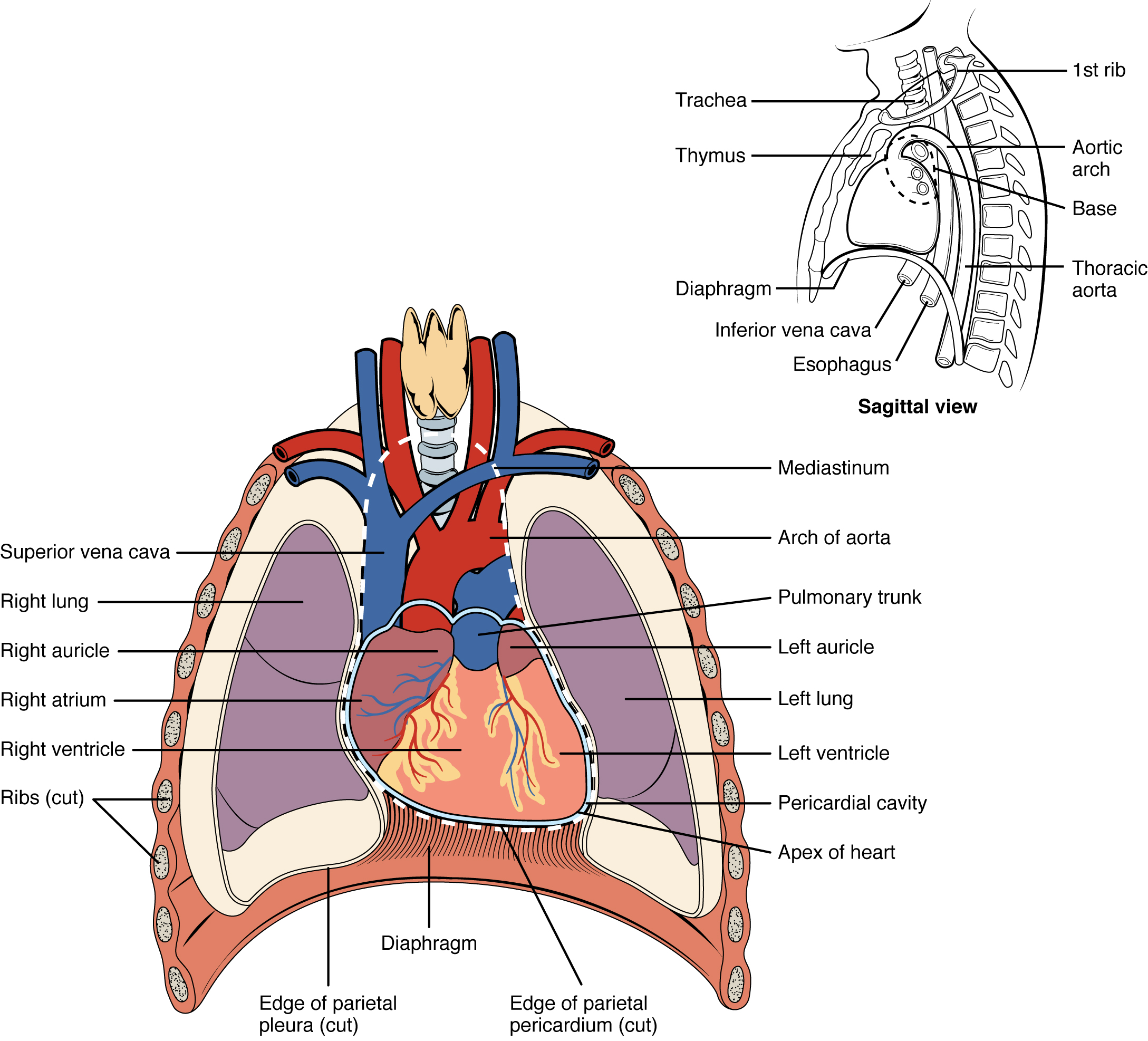
Gross Anatomy & Location
<ul><li><strong>Location:</strong> Mediastinum within the thoracic cavity, resting on the diaphragm.</li><li><strong>Orientation:</strong> Apex points anteriorly, inferiorly, and to the left; Base faces posteriorly.</li><li><strong>Pericardium:</strong><ul><li><em>Fibrous Pericardium:</em> Tough outer layer anchors heart to surrounding structures.</li><li><em>Serous Pericardium:</em> Parietal and Visceral (Epicardium) layers separated by fluid-filled pericardial cavity.</li></ul></li></ul>
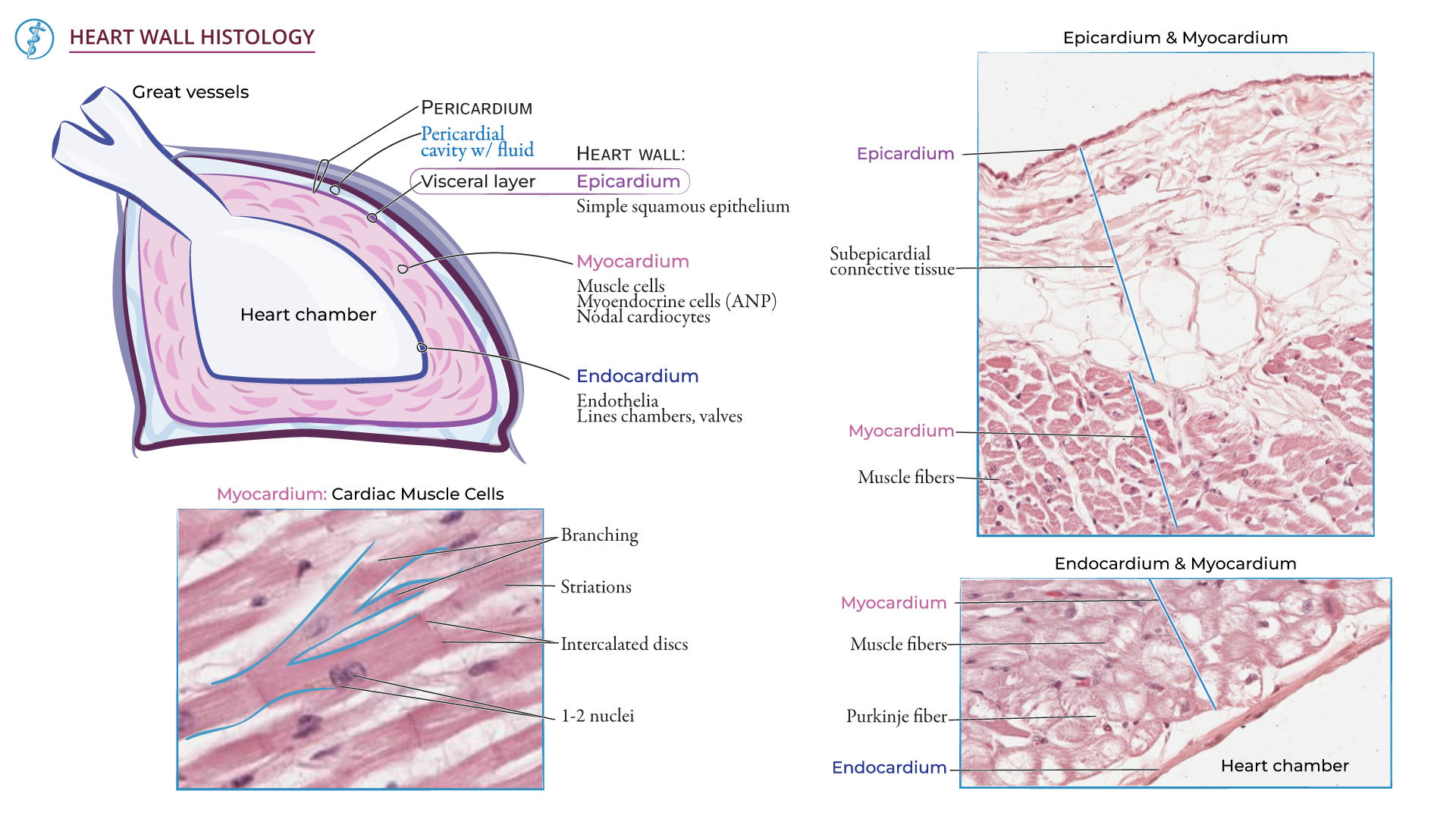
Layers of the Heart Wall
<ul><li><strong>Epicardium (Outer):</strong> Visceral layer of serous pericardium; mesothelium and connective tissue (often contains fat).</li><li><strong>Myocardium (Middle):</strong> Thickest layer; cardiac muscle arranged in spiral bundles; contains intercalated discs for electrical coupling.</li><li><strong>Endocardium (Inner):</strong> Smooth endothelium resting on a thin connective tissue layer; continuous with blood vessel lining to reduce friction.</li></ul>
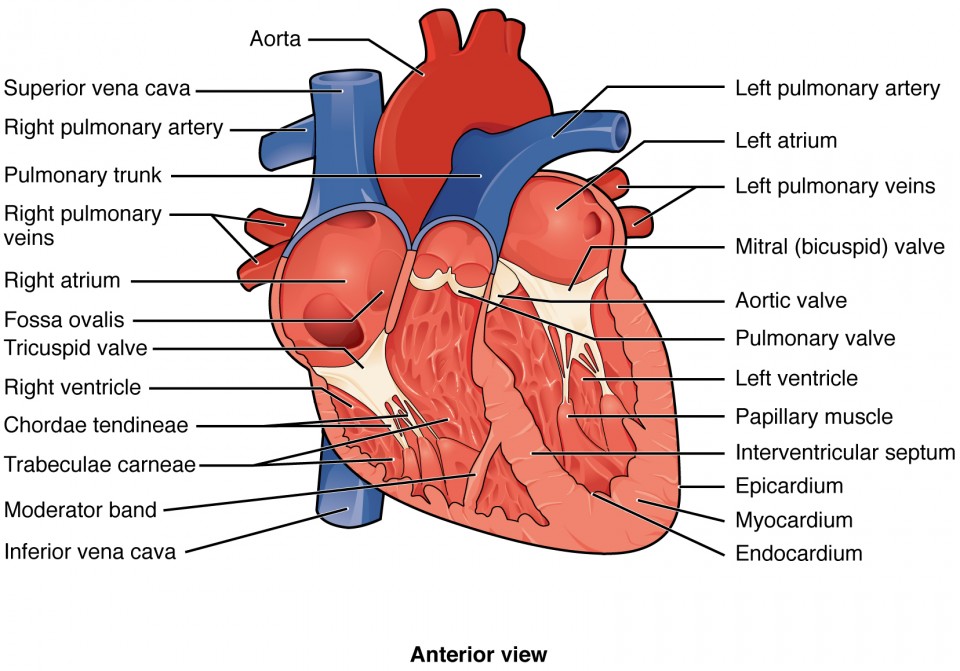
The Four Chambers
<ul><li><strong>Atria (Receiving Chambers):</strong><ul><li>Thin-walled, separated by <em>Interatrial Septum</em> (Fossa Ovalis).</li><li>Contains Pectinate muscles (mainly in right atrium) and Auricles (ear-like flaps).</li></ul></li><li><strong>Ventricles (Pumping Chambers):</strong><ul><li>Thick-walled, separated by <em>Interventricular Septum</em>.</li><li>Lined with <em>Trabeculae Carneae</em> ridges.</li><li><strong>Left Ventricle:</strong> Significantly thicker wall to pump against systemic pressure.</li></ul></li></ul>
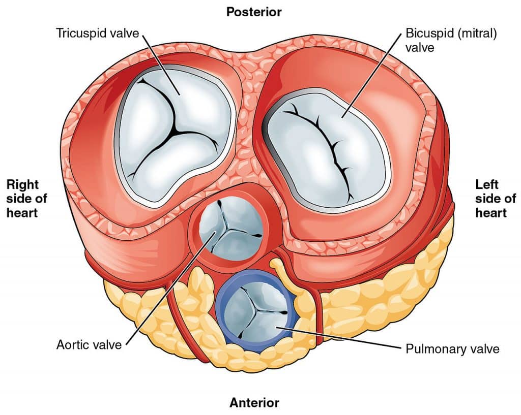
Heart Valves: Structural Gates
<ul><li><strong>Atrioventricular (AV) Valves:</strong><ul><li><em>Tricuspid (Right)</em> & <em>Mitral/Bicuspid (Left)</em>.</li><li>Anchored by <strong>Chordae Tendineae</strong> ("heart strings") to <strong>Papillary Muscles</strong> protruding from ventricle walls.</li></ul></li><li><strong>Semilunar (SL) Valves:</strong><ul><li><em>Pulmonary</em> & <em>Aortic</em> valves.</li><li>Three pocket-like cusps; no chordae/papillary muscles.</li><li>Open under pressure; close to prevent backflow into ventricles.</li></ul></li></ul>
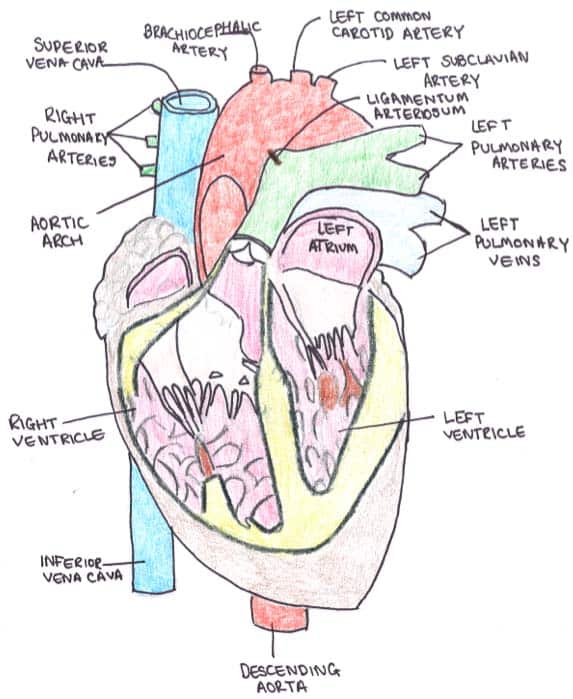
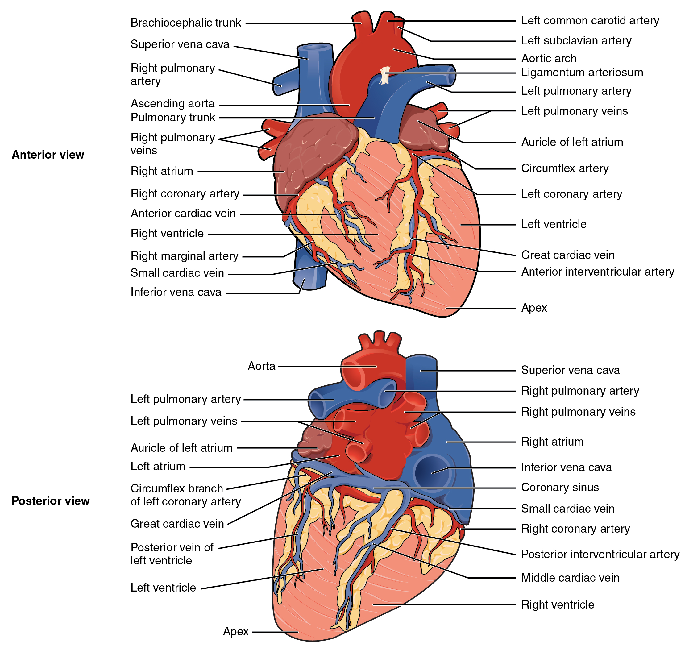
Great Vessels of the Heart
<ul><li><strong>Vena Cavae:</strong> Superior and Inferior; enter Right Atrium (deoxygenated blood).</li><li><strong>Pulmonary Trunk:</strong> Exits Right Ventricle; bifurcates into Left/Right Pulmonary Arteries.</li><li><strong>Pulmonary Veins:</strong> Four vessels (2 left, 2 right) entering Left Atrium (oxygenated blood).</li><li><strong>Aorta:</strong> Largest artery; exits Left Ventricle. Segments: Ascending Aorta, Aortic Arch, Descending Aorta.</li><li><strong>Ligamentum Arteriosum:</strong> Connective tissue remnant between aorta and pulmonary trunk.</li></ul>
Coronary Circulation
<ul><li><strong>Coronary Arteries:</strong> Arise from the base of the aorta (coronary sinuses).<ul><li><em>Left CA:</em> Divides into Anterior Interventricular (LAD) and Circumflex arteries.</li><li><em>Right CA:</em> Supplies Marginal artery and Posterior Interventricular artery.</li></ul></li><li><strong>Cardiac Veins:</strong> Great, Middle, and Small cardiac veins drain myocardium.</li><li><strong>Coronary Sinus:</strong> Large vein on posterior heart; collects venous blood and empties into Right Atrium.</li></ul>
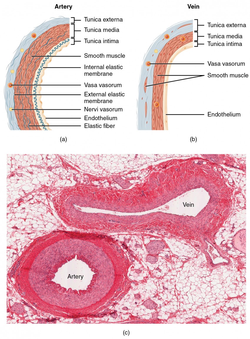
Vessel Wall Structure: Tissue Composition
<div style='margin-bottom:30px;'>Structure dictates function:<ul><li><strong>Aorta:</strong> High <em>elastic</em> content absorbs pressure pulses.</li><li><strong>Muscular Artery:</strong> High <em>smooth muscle</em> regulates flow distribution.</li><li><strong>Vein:</strong> High <em>fibrous</em> content; less muscle/elastic.</li></ul></div>
Arteries vs. Veins: Structural Differences
<table style='width:100%; border-collapse:collapse; font-size:26px;'><thead><tr style='background:#eee;'><th style='padding:15px; text-align:left;'>Feature</th><th style='padding:15px; text-align:left; color:#c0392b;'>Arteries</th><th style='padding:15px; text-align:left; color:#2980b9;'>Veins</th></tr></thead><tbody><tr><td style='padding:15px; border-bottom:1px solid #ddd;'><strong>Lumen</strong></td><td style='padding:15px; border-bottom:1px solid #ddd;'>Round, stays open</td><td style='padding:15px; border-bottom:1px solid #ddd;'>Irregular, collapsed</td></tr><tr><td style='padding:15px; border-bottom:1px solid #ddd;'><strong>Tunica Media</strong></td><td style='padding:15px; border-bottom:1px solid #ddd;'>Thickest layer (Muscle/Elastic)</td><td style='padding:15px; border-bottom:1px solid #ddd;'>Thin</td></tr><tr><td style='padding:15px; border-bottom:1px solid #ddd;'><strong>Tunica Externa</strong></td><td style='padding:15px; border-bottom:1px solid #ddd;'>Thinner than media</td><td style='padding:15px; border-bottom:1px solid #ddd;'>Thickest layer (Collagen)</td></tr><tr><td style='padding:15px; border-bottom:1px solid #ddd;'><strong>Valves</strong></td><td style='padding:15px; border-bottom:1px solid #ddd;'>Absent</td><td style='padding:15px; border-bottom:1px solid #ddd;'>Present (in limbs)</td></tr></tbody></table>
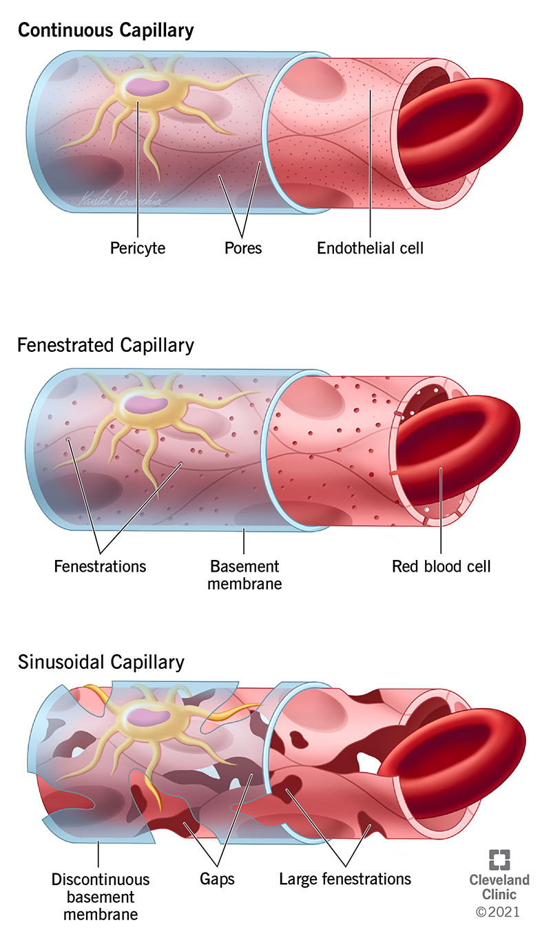
Microcirculation Structure
<ul><li><strong>Arterioles:</strong> Smallest arteries; Tunica media is mostly smooth muscle (regulates resistance).</li><li><strong>Capillaries:</strong><ul><li>Microscopic tubes comprising only <em>Endothelium</em> and <em>Basement Membrane</em>.</li><li><strong>Continuous:</strong> Tight junctions (Skin, Muscle).</li><li><strong>Fenestrated:</strong> Pores for permeability (Kidney, Small Intestine).</li><li><strong>Sinusoids:</strong> Large gaps (Liver, Spleen).</li></ul></li><li><strong>Venules:</strong> Porous post-capillary vessels; unite to form veins.</li></ul>
- heart-anatomy
- cardiovascular-system
- pre-med
- coronary-circulation
- heart-valves
- vessel-histology