


Fins & Frameworks: Fish Appendicular Skeleton Anatomy
Explore the structural anatomy and evolution of fish fins and girdles. Learn about the differences between bony and cartilaginous skeletal systems.
ICHTHYOLOGY · VERTEBRATE ANATOMY
ENDOSKELETON
IN FISHES
Appendicular Skeleton
Structure · Girdles · Fins · Evolution
Zoology | Fish Anatomy
03
INTRODUCTION TO FISH ENDOSKELETON
What is an Endoskeleton?
An endoskeleton is an internal framework of bones or cartilage that supports and protects the body of an organism. In fishes, it forms the structural foundation for movement, protection, and organ support.
Types of Fish Skeleton
Bony Fishes (Osteichthyes) — ossified endoskeleton
Cartilaginous Fishes (Chondrichthyes) — cartilaginous endoskeleton
Divisions of Fish Endoskeleton
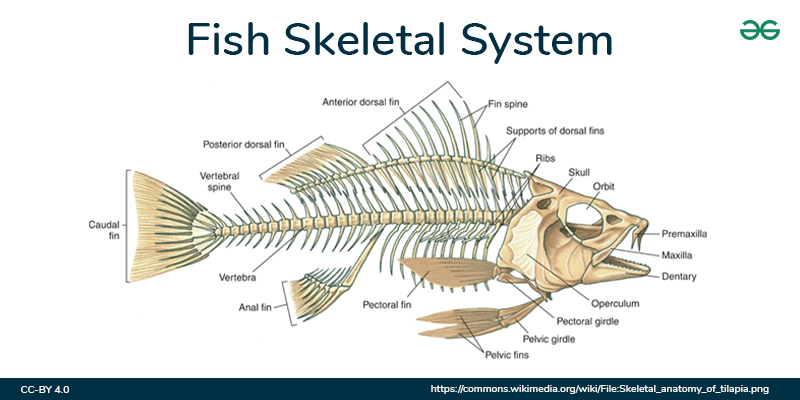
AXIAL SKELETON
APPENDICULAR SKELETON
VISCERAL SKELETON
Ichthyology | Vertebrate Anatomy
04
APPENDICULAR SKELETON — OVERVIEW
Definition
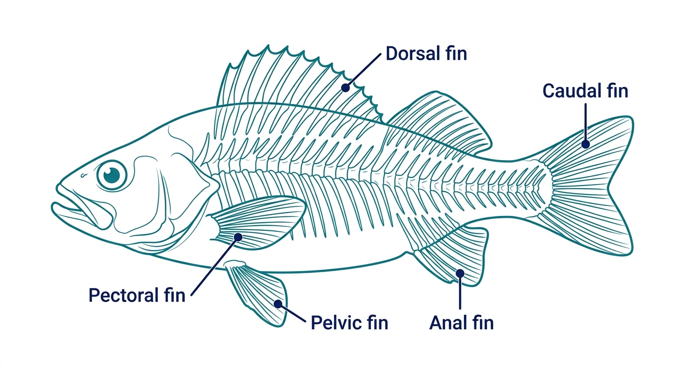
The appendicular skeleton consists of all paired fins and their supporting girdles. It is the fish equivalent of the limb skeleton in tetrapods.
Key Function:
Locomotion, balance, steering and mating displays
Components
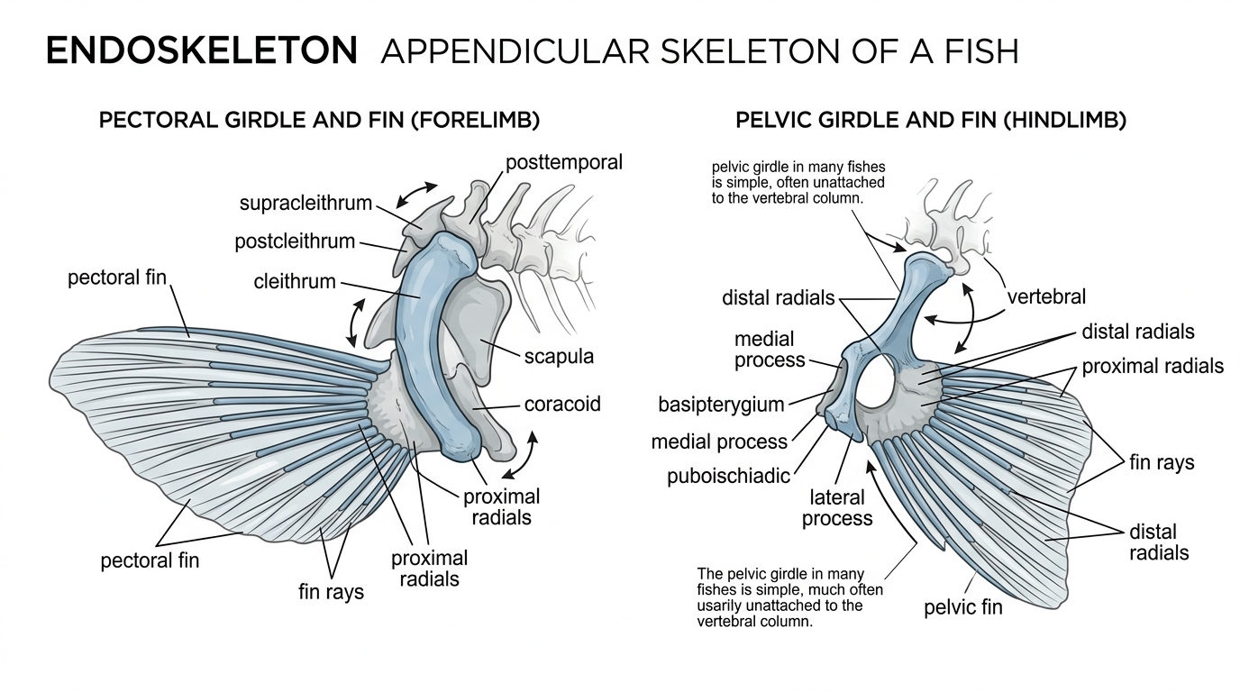
Pectoral Girdle
Pectoral Fins
Pelvic Girdle
Pelvic Fins
Paired fins = homologous to tetrapod limbs
PAIRED APPENDAGES
Zoology | Fish Anatomy
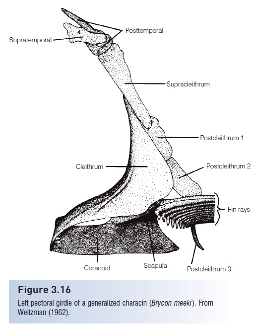
PECTORAL GIRDLE
Structure, Composition & Bone Elements
The pectoral girdle anchors the pectoral fins to the body. In bony fishes, it attaches to the skull rather than the vertebral column.
Endochondral Bones
Dermal (Intramembranous) Bones
In cartilaginous fishes (sharks), the pectoral girdle is a single large
SCAPULOCORACOID
with scapular process, suprascapula, and coracoid bar.
Fig: Left Pectoral Girdle of a generalized bony fish
ICHTHYOLOGY | VERTEBRATE ANATOMY
06
PECTORAL FIN ANATOMY
Skeletal Components & Radial Organization
CLEITHRUM
Crescent-shaped dermal bone
SCAPULA + CORACOID
Endochondral bones
MESOCORACOID
Connects cleithrum (lost in derived teleosts)
PROXIMAL RADIALS (PR)
Elongated bones, typically 4
DISTAL RADIALS (DR)
Granular connecting bones
FIN RAYS (FR)
Rod-shaped dermal terminal bones
4-Basal Rule: Most teleosts have 4 proximal radials
Mesocoracoid absent in advanced teleosts
Fin ray connections: 1-to-many (basal) vs 1-to-1 (derived)
07
PROXIMAL RADIAL MORPHOLOGY
Evolutionary Shape Variations Across Teleosts
BASAL TELEOSTS
RECTANGULAR-TYPE
Adjacent along the long side. Found in basal teleosts.
Osteoglossomorpha, Otomorpha, Protacanthopterygii
DERIVED TELEOSTS
HOURGLASS-TYPE
Adjacent only at distal and proximal tips. Wide space in the middle.
Paracanthopterygii and Acanthopterygii
ADVANCED TELEOSTS
DUMBBELL-TYPE
Tightly adjacent with holes in mid-portion. Hollowed after chondrification.
Syngnatharia, Gobiaria, Eupercaria
Evolutionary Progression →
Pectoral Girdle Comparison
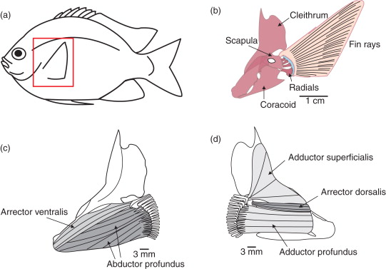
PELVIC GIRDLE
Structure, Attachment & Composition
Unlike the pectoral girdle, the pelvic girdle in fishes is <strong>NOT</strong> attached to the vertebral column — it remains <strong>FREE</strong> within the body wall.
The pelvic girdle arose from pterygophores and is fundamentally simpler than the tetrapod pelvis (which has pubis, ilium & ischium attached to vertebral column).
Fig: Fish Pectoral & Pelvic Girdles comparison
PELVIC FIN ANATOMY
Skeletal Structure & Positional Variations
Each pelvic fin supported by fin rays (lepidotrichia) and radials (dermactidia)
In <em>Labeo rohita</em>: 9 lepidotrichia + 3 radials
First 2 radials → 2 fin rays each; 3rd radial → 3 fin rays
Articulate with posterior border of basipterygium
ABDOMINAL POSITION
Pelvic fins located ventrally, anterior to anal fin
Soft-rayed teleosts (e.g., salmon, herring)
PRIMITIVE POSITION
THORACIC POSITION
Directly below the pectoral fins
Spiny-rayed fishes (e.g., perch, bass)
ADVANCED POSITION
JUGULAR POSITION
Anterior to pectoral fins; may attach to pectoral girdle
Cusk-eels, cods
SPECIALIZED
<strong>Pelvic girdle entirely LOST in:</strong> eels (Anguilliformes), puffers, and some needlefishes
10
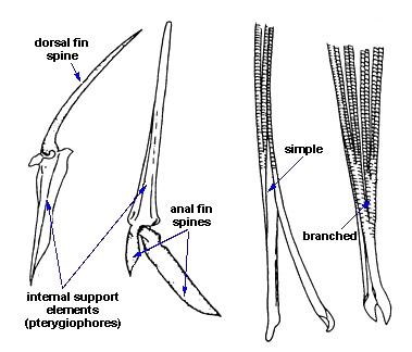
FIN RAY TYPES
Spines & Soft Rays — Structure & Function
Fig: Comparison of fin spines and soft rays
SPINES
Simple, Unbranched, Unsegmented
<b>Hard, stiff, pointed</b> structures
Single piece of bone — <b>not paired</b>
Found in more evolutionarily <span style="color:#ff9800; font-weight:700;">ADVANCED</span> fishes
Example: First dorsal fin rays in perch
Function: <b>Defense and support</b>
SOFT RAYS
Branched, Segmented, Flexible
Composed of two mirrored halves <b>(paired)</b>
<b>Flexible, branched</b> at tips
Found in <b>primitive and derived</b> fishes
Example: Pectoral and pelvic fins
Function: <b>Fine locomotor control</b>
<span style="color:#ffffff; font-weight:700;">Lepidotrichia</span> = dermal fin rays | <span style="color:#ffffff; font-weight:700;">Actinotrichia</span> = ceratotrichia in cartilaginous fishes
11
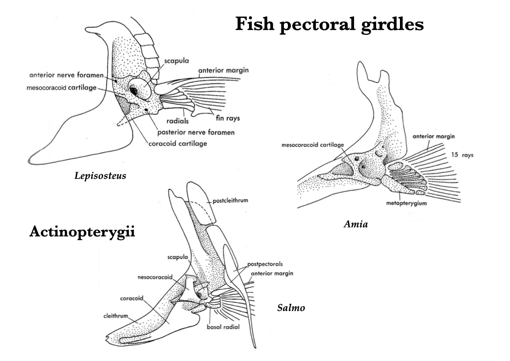
MESOCORACOID — EVOLUTIONARY LOSS
A Key Innovation in Teleost Evolution
The mesocoracoid is a bone present in all basal actinopterygians (sturgeon, paddlefish, gar, bowfin). Its selective LOSS in derived teleosts is a major evolutionary event.
BASAL ACTINOPTERYGIANS
✓ Mesocoracoid PRESENT
Sturgeon, Paddlefish, Gar, Bowfin
Pectoral fins positioned VENTRALLY
Fins protrude laterally from ventral side
BASAL TELEOSTS
✓ Mesocoracoid PRESENT
Osteoglossomorpha, Otomorpha
Pectoral fins still ventral
Rectangular proximal radials
DERIVED TELEOSTS
✗ Mesocoracoid LOST
Paracanthopterygii, Acanthopterygii
Pectoral fins repositioned LATERALLY
Hourglass/dumbbell radials
Better fin control and maneuverability
🔬 In zebrafish larvae (8.2–8.9 mm body length), pectoral fins shift from lateral → ventral positioning, coinciding with mesocoracoid cartilage formation — suggesting developmental linkage.
Fig: Evolution of pectoral girdles across fish groups
EVOLUTIONARY ADAPTATIONS
Special Cases & Remarkable Modifications
These diverse modifications demonstrate the remarkable plasticity of the appendicular skeleton across 30,000+ fish species
13
BONY FISH vs CARTILAGINOUS FISH
Appendicular Skeleton Comparison
BONY FISHES (Osteichthyes)
🦴 OSSIFIED SKELETON
Composed of both endochondral AND dermal bones; cleithrum always present
Attaches to SKULL (not vertebral column)
Endochondral only; puboischiac bar fuses left & right halves
Present; single element
Lepidotrichia (bony, segmented, often branched)
Present in basal; LOST in derived teleosts
Salmon, Perch, Cod, Rohu
CARTILAGINOUS FISHES (Chondrichthyes)
💎 CARTILAGINOUS SKELETON
Single large SCAPULOCORACOID cartilage (scapula + coracoid fused)
Free; NOT attached to skull or vertebral column
Single acetabular cartilage with iliac process
Cartilaginous basipterygium
Ceratotrichia (unsegmented, flexible protein fibers)
ABSENT throughout
Sharks, Rays, Skates
SUMMARY // Bony fishes possess an ossified appendicular skeleton attached to the skull, while cartilaginous fishes have a free connective cartilage structure.
KEY TAKEAWAYS
Endoskeleton in Fishes: Appendicular Skeleton
The <b>appendicular skeleton</b> = paired fins + girdles that support them
<b>Pectoral girdle</b> attaches to skull (not spine) in bony fishes; contains both endochondral and dermal bones
<b>Pelvic girdle</b> is FREE from the axial skeleton; composed of endochondral bone only
<b>Proximal radials</b> follow the 4-basal rule; shaped as rectangular → hourglass → dumbbell through evolution
<b>Loss of mesocoracoid</b> in derived teleosts enabled lateral repositioning of pectoral fins — key innovation
<b>Pelvic fin position varies:</b> abdominal (primitive) → thoracic → jugular (advanced/specialized)
The appendicular skeleton reflects 400+ million years of fish evolution and adaptation
- ichthyology
- fish-anatomy
- zoology
- vertebrate-anatomy
- evolution
- marine-biology