Advances in Artificial Skin: 3D Bioprinting & E-Skin
Explore the future of artificial skin, from 3D bioprinting living tissues to electronic E-skin sensors for robotics and prosthetics.
Artificial Skin Technologies
From 3D Bioprinting to Electronic Skin Sensors
Defining the Scope: Biological vs. Synthetic
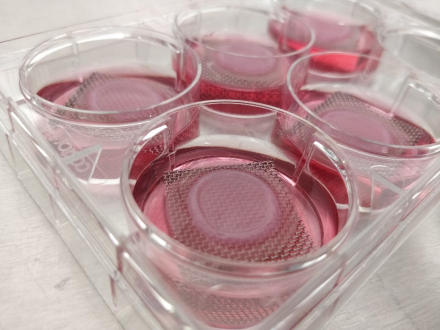
Tissue Engineered Skin
Living cells (primary fibroblasts, keratinocytes, stem cells) seeded in collagen/alginate scaffolds. Focus: chronic wound healing and burn regeneration.
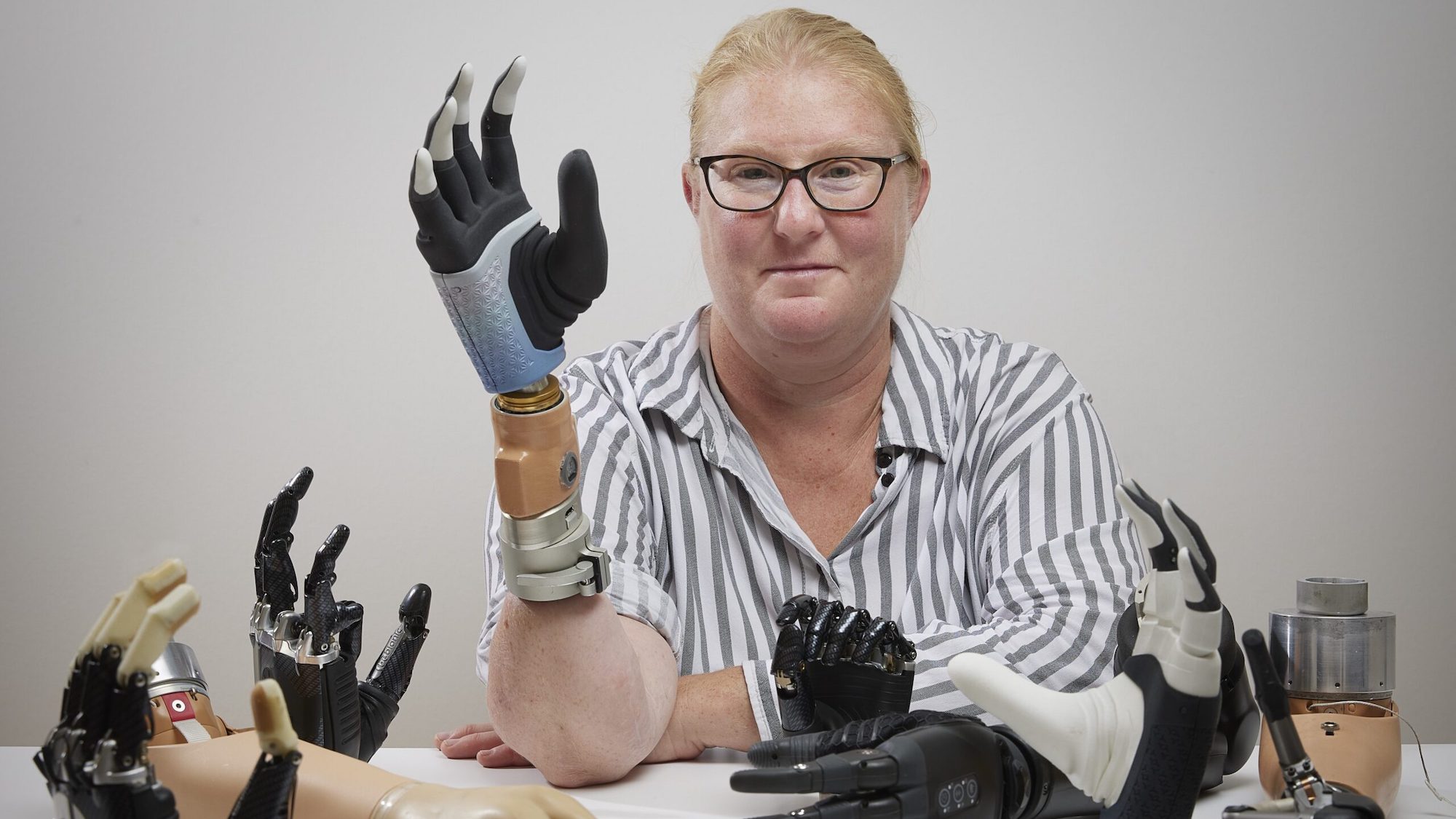
Electronic Skin (E-Skin)
Polymers (PDMS, Polyimide) embedded with piezoelectric/capacitive sensors. Focus: robotic tactile sensing, prosthetics, and wearable health monitoring.
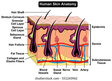
The Biological Target: Native Architecture
Successful engineering requires mimicking the tri-layered structure:
Epidermis: Stratum corneum barrier, keratinocyte differentiation, tight junctions prevent fluid loss.
Dermis: ECM rich in Type I/III collagen, mechanoreceptors (Meissner/Pacinian), vascular networks.
Hypodermis: Adipocyte lobules for energy storage, thermal insulation, and mechanical shock absorption.
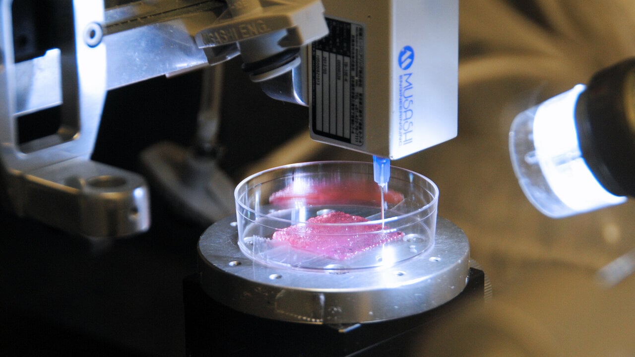
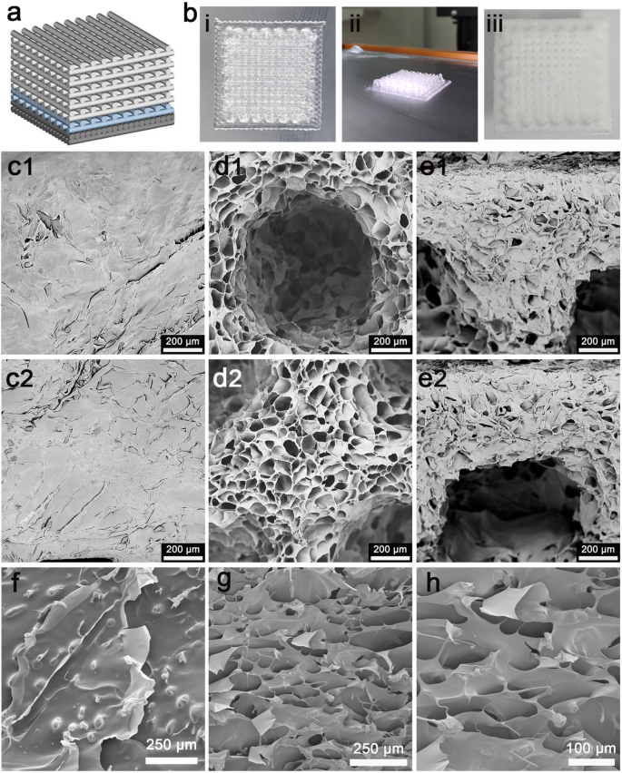
3D Bioprinting Fabrication
Extrusion-based bioprinting uses 'bioinks'—shear-thinning hydrogels (GelMA, Alginate) encapsulating cells. Key parameters include nozzle diameter (200-400µm), printing pressure, and crosslinking mechanisms (UV, enzymatic) to ensure >85% cell viability.
Targeting Engineering Constraints
Target Thickness for Engineered Layers (mm)
A critical challenge in tissue engineering is creating layers of appropriate thickness while ensuring vascular perfusion. Grafts thicker than 400μm require internal vascular networks to survive.
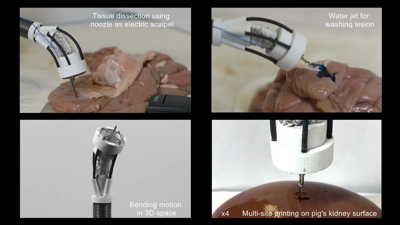
In Situ Bioprinting
Direct-to-wound printing utilizes robotic arms to scan wound geometry.
Deposits fibrin-based bioinks containing autologous cells.
Advantages: Precise fit for irregular burn wounds, reduced surgery time.
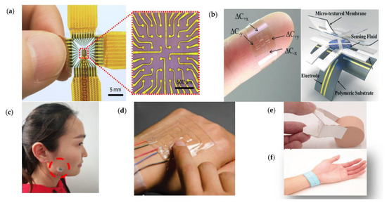
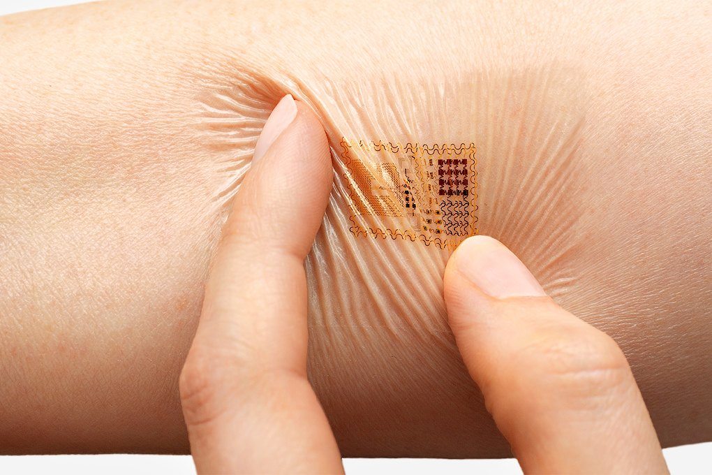
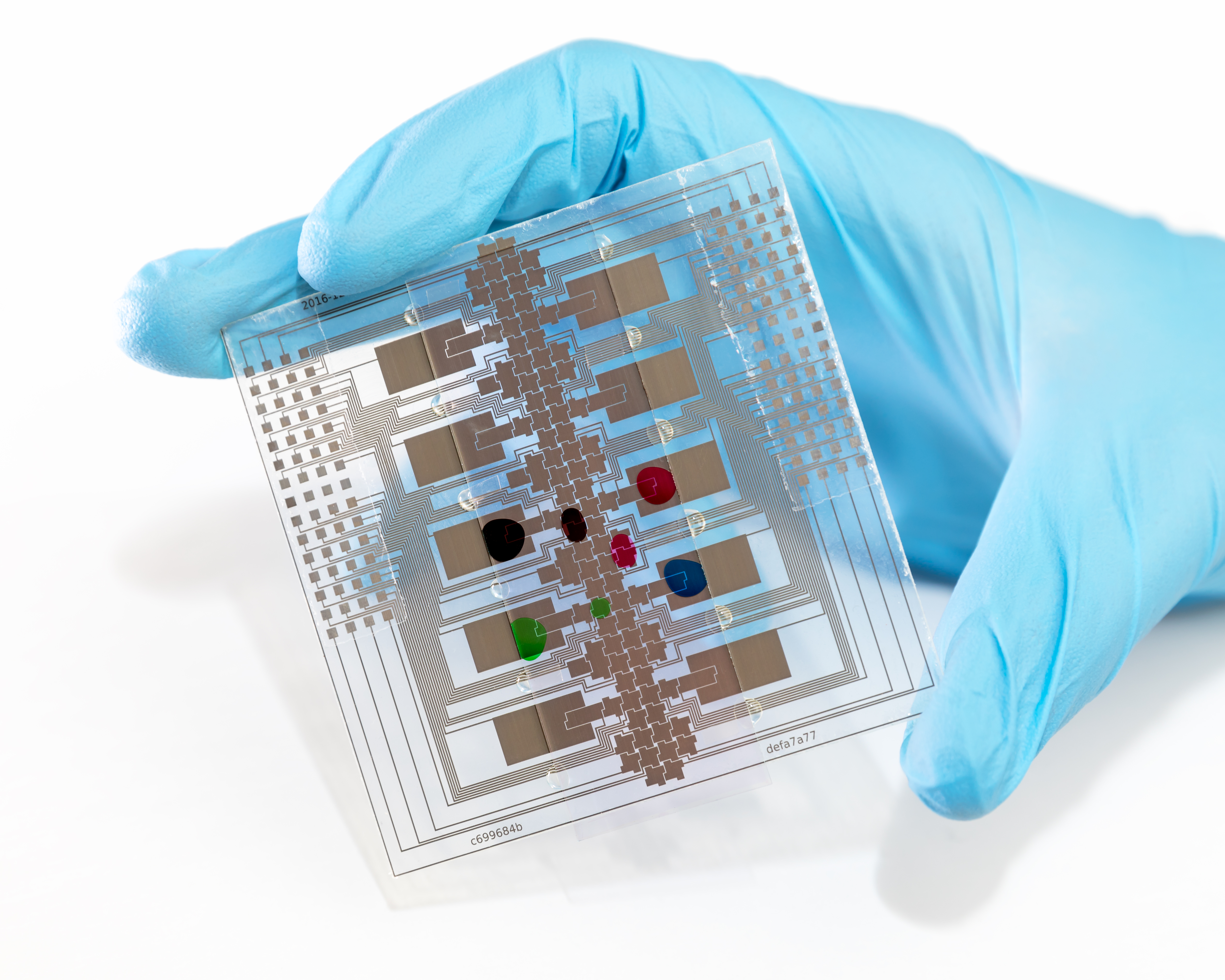
Electronic Skin (E-Skin) Structures
Synthesizing touch: Mimicking mechanoreceptors through flexible electronics.
Piezoresistive: Conductive elastomers change resistance upon deformation. Ideal for dynamic pressure monitoring (1-100 kPa range).
Capacitive Arrays: Dielectric layers sandwiched between electrodes. High sensitivity (<1 Pa) for proximity and light touch detection.
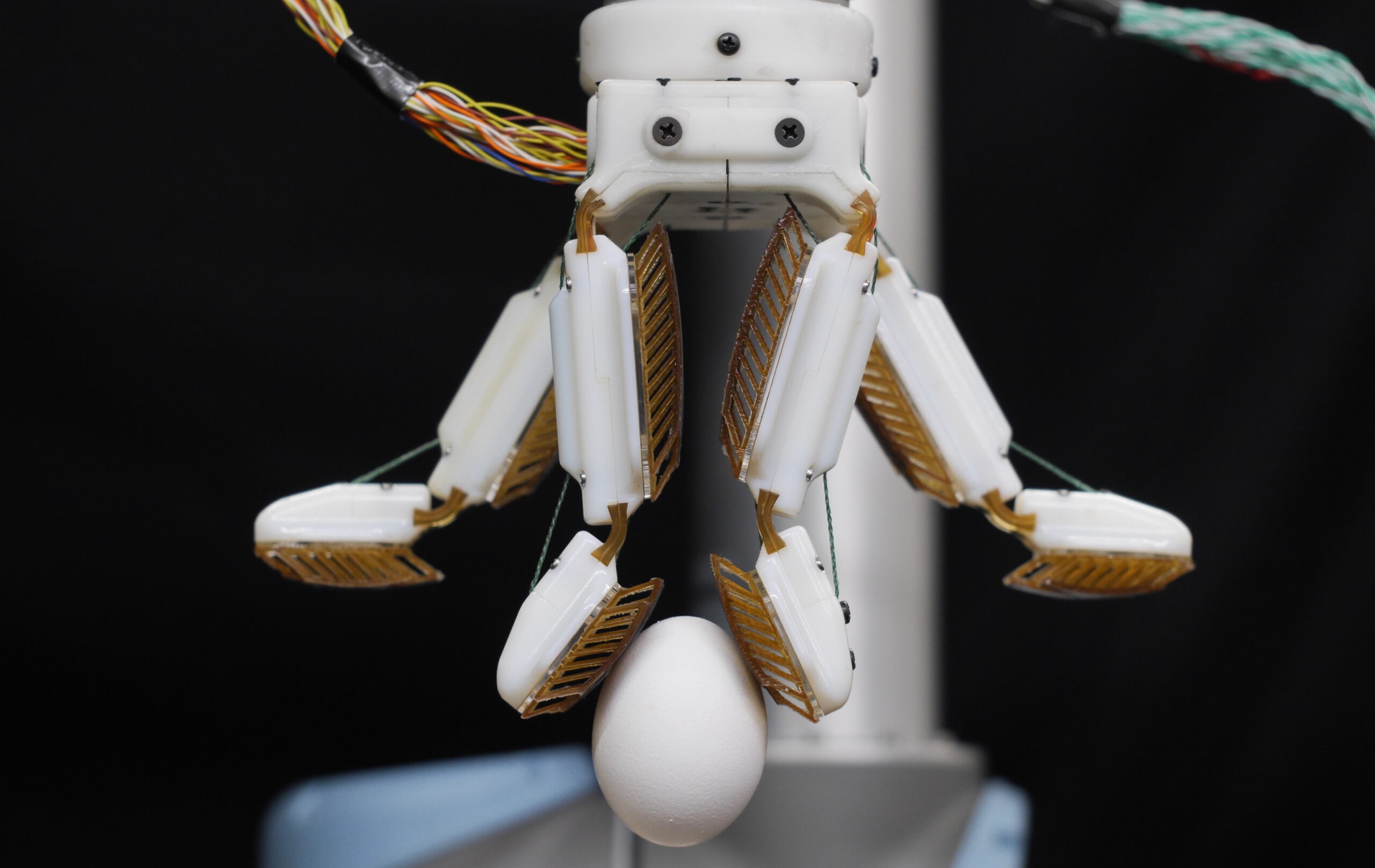
Applications in Robotics & Prosthetics
Advanced e-skin mimics the somatosensory cortex feedback loop using multi-modal sensor arrays (strain, thermal, pressure). This enables robotic grippers to distinguish texture and stiffness, preventing damage to delicate objects.
Clinical Advantages of Bioprinting
Eliminates Donor Site Need
Reduces morbidity: Autografts have a 5-10% complication rate. Bioprinting eliminates secondary wound sites.
Customization
Patient-specific cell matching reduces rejection; pigment matching possible.
Speed & Scalability
High-throughput: Specialized bioprinters can deposit 10-50 cm²/hour, significantly faster than traditional manual epithelial sheet culture.
Future Challenges
Key Hurdles to Commercialization
Vascularization
Diffusion limit of oxygen is ~200μm. Thick tissues require sacrificial channels or angiogenesis factors to prevent core necrosis.
Cost & Regulation
High CAPEX for GMP bioprinters; complex 510(k)/PMA regulatory pathways due to combination product classification (biologic/device).
Summary & Vision
The convergence of 3D bioprinting and materials science is moving us towards 'smart skin'—tissues that not only heal wounds but restore sensation and integrate seamlessly with both biology and robotics.
- 3d-bioprinting
- electronic-skin
- tissue-engineering
- robotics
- biomedical-engineering
- medical-technology
- prosthetics