Haemorrhagic Shock: Pathophysiology and ATLS Management
Master the classification, pathophysiology, and life-saving management of haemorrhagic shock with this comprehensive guide based on ATLS guidelines.
Haemorrhagic Shock
Pathophysiology, Classification & Management
A Comprehensive Medical Overview
Contents
Definition & Overview
Aetiology & Causes
Pathophysiology
Classification (Classes I–IV)
Clinical Features & Diagnosis
Complications
Management & Treatment
01 / Definition
What is Haemorrhagic Shock?
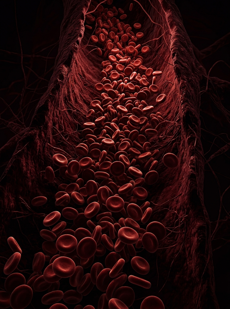
Haemorrhagic shock is a form of hypovolaemic shock resulting from acute blood loss, leading to inadequate tissue perfusion and cellular hypoxia.
<strong>Also known as:</strong> Class IV Hypovolaemic Shock
Accounts for ~30–40% of trauma-related deaths worldwide
Most common cause of preventable death in trauma
Rapid recognition and intervention is life-saving
Source: Advanced Trauma Life Support (ATLS) Guidelines
Aetiology & Causes
Trauma (most common)
Blunt trauma
Penetrating injuries
Crush injuries
Gastrointestinal
Peptic ulcer bleeding
Variceal haemorrhage
Mallory-Weiss tear
Obstetric
Postpartum haemorrhage
Placenta praevia
Ectopic pregnancy rupture
Surgical
Intraoperative haemorrhage
Post-surgical bleeding
Vascular
Aortic aneurysm rupture
Arterial laceration
Iatrogenic
Anticoagulant overdose
Procedural complications
Pathophysiology
Acute Blood Loss
↓ Circulating Blood Volume
↓ Venous Return (Preload)
↓ Cardiac Output
↓ Mean Arterial Pressure
Baroreceptor Activation
Sympathetic Nervous System Activation
Vasoconstriction + ↑ Heart Rate
↓ Tissue Perfusion
Cellular Hypoxia
Anaerobic Metabolism
Lactic Acidosis
Multi-Organ Dysfunction
Compensatory mechanisms (Steps 6–8) may temporarily maintain BP — masking severity.
Classification (ATLS)
American College of Surgeons — Advanced Trauma Life Support
CLASS I
(Mild)
CLASS II
(Moderate)
CLASS III
(Severe)
CLASS IV
(Life-threatening)
Blood Loss
Heart Rate
Blood Pressure
Pulse Pressure
Respiratory Rate
Urine Output
Mental Status
Up to 750 mL / <15%
<100 bpm
Normal
Normal
14–20 / min
>30 mL/hr
Slightly anxious
750–1500 mL / 15–30%
100–120 bpm
Normal
Decreased
20–30 / min
20–30 mL/hr
Mildly anxious
1500–2000 mL / 30–40%
120–140 bpm
Decreased
Decreased
30–40 / min
5–15 mL/hr
Anxious, confused
>2000 mL / >40%
>140 bpm
Decreased
Decreased
>35 / min
Negligible
Confused, lethargic
Clinical Features
& Diagnosis
Complications
Acute Kidney Injury (AKI)
Renal hypoperfusion → tubular necrosis → oliguria/anuria
Acute Respiratory Distress Syndrome (ARDS)
Inflammatory response → pulmonary oedema → hypoxaemia
Disseminated Intravascular Coagulation (DIC)
Consumption of clotting factors → uncontrolled bleeding
Multi-Organ Dysfunction Syndrome (MODS)
Cascading organ failure (liver, kidneys, heart, lungs)
Ischaemic Heart Disease / Cardiac Arrest
Myocardial hypoperfusion → arrhythmias → arrest
Death
Without timely intervention, mortality can exceed 40%
Management & Treatment
The Goal: Stop Bleeding + Restore Perfusion
Initial Resuscitation
Haemorrhage Control
Monitoring & Supportive Care
<div style="display: flex; align-items: flex-start;"><div style="color: #8B0000; font-size: 26px; margin-right: 14px; line-height: 1.1;">•</div><div style="font-size: 22px; color: #2C3E50; line-height: 1.4; font-weight: 400;">Primary ABCDE survey</div></div><div style="display: flex; align-items: flex-start;"><div style="color: #8B0000; font-size: 26px; margin-right: 14px; line-height: 1.1;">•</div><div style="font-size: 22px; color: #2C3E50; line-height: 1.4; font-weight: 400;">High-flow O₂ via non-rebreather mask</div></div><div style="display: flex; align-items: flex-start;"><div style="color: #8B0000; font-size: 26px; margin-right: 14px; line-height: 1.1;">•</div><div style="font-size: 22px; color: #2C3E50; line-height: 1.4; font-weight: 400;">Two large-bore IV access (14–16G)</div></div><div style="display: flex; align-items: flex-start;"><div style="color: #8B0000; font-size: 26px; margin-right: 14px; line-height: 1.1;">•</div><div style="font-size: 22px; color: #2C3E50; line-height: 1.4; font-weight: 400;">Warm IV fluids — Hartmann's / Normal Saline (cautious use)</div></div><div style="display: flex; align-items: flex-start;"><div style="color: #8B0000; font-size: 26px; margin-right: 14px; line-height: 1.1;">•</div><div style="font-size: 22px; color: #2C3E50; line-height: 1.4; font-weight: 400;">Blood products: O-negative pRBCs in emergencies</div></div><div style="display: flex; align-items: flex-start;"><div style="color: #8B0000; font-size: 26px; margin-right: 14px; line-height: 1.1;">•</div><div style="font-size: 22px; color: #2C3E50; line-height: 1.4; font-weight: 400;">Massive Transfusion Protocol (MTP): 1:1:1 ratio (RBC:FFP:Platelets)</div></div><div style="display: flex; align-items: flex-start;"><div style="color: #8B0000; font-size: 26px; margin-right: 14px; line-height: 1.1;">•</div><div style="font-size: 22px; color: #2C3E50; line-height: 1.4; font-weight: 400;">Tranexamic Acid (TXA) within 3 hours of injury</div></div>
<div style="display: flex; align-items: flex-start;"><div style="color: #4A0010; font-size: 26px; margin-right: 14px; line-height: 1.1;">•</div><div style="font-size: 22px; color: #2C3E50; line-height: 1.4; font-weight: 400;">Direct pressure / tourniquet for external bleeding</div></div><div style="display: flex; align-items: flex-start;"><div style="color: #4A0010; font-size: 26px; margin-right: 14px; line-height: 1.1;">•</div><div style="font-size: 22px; color: #2C3E50; line-height: 1.4; font-weight: 400;">Surgical haemostasis — damage control surgery</div></div><div style="display: flex; align-items: flex-start;"><div style="color: #4A0010; font-size: 26px; margin-right: 14px; line-height: 1.1;">•</div><div style="font-size: 22px; color: #2C3E50; line-height: 1.4; font-weight: 400;">Interventional radiology — angioembolisation</div></div><div style="display: flex; align-items: flex-start;"><div style="color: #4A0010; font-size: 26px; margin-right: 14px; line-height: 1.1;">•</div><div style="font-size: 22px; color: #2C3E50; line-height: 1.4; font-weight: 400;">Wound packing with haemostatic agents</div></div><div style="display: flex; align-items: flex-start;"><div style="color: #4A0010; font-size: 26px; margin-right: 14px; line-height: 1.1;">•</div><div style="font-size: 22px; color: #2C3E50; line-height: 1.4; font-weight: 400;">Pelvic binder for pelvic fractures</div></div><div style="display: flex; align-items: flex-start;"><div style="color: #4A0010; font-size: 26px; margin-right: 14px; line-height: 1.1;">•</div><div style="font-size: 22px; color: #2C3E50; line-height: 1.4; font-weight: 400;">Avoid hypothermia, acidosis, coagulopathy (lethal triad)</div></div>
<div style="display: flex; align-items: flex-start;"><div style="color: #1A1A1A; font-size: 26px; margin-right: 14px; line-height: 1.1;">•</div><div style="font-size: 22px; color: #2C3E50; line-height: 1.4; font-weight: 400;">Urinary catheter — urine output target >0.5 mL/kg/hr</div></div><div style="display: flex; align-items: flex-start;"><div style="color: #1A1A1A; font-size: 26px; margin-right: 14px; line-height: 1.1;">•</div><div style="font-size: 22px; color: #2C3E50; line-height: 1.4; font-weight: 400;">Arterial line for continuous BP monitoring</div></div><div style="display: flex; align-items: flex-start;"><div style="color: #1A1A1A; font-size: 26px; margin-right: 14px; line-height: 1.1;">•</div><div style="font-size: 22px; color: #2C3E50; line-height: 1.4; font-weight: 400;">Central venous access (CVP monitoring)</div></div><div style="display: flex; align-items: flex-start;"><div style="color: #1A1A1A; font-size: 26px; margin-right: 14px; line-height: 1.1;">•</div><div style="font-size: 22px; color: #2C3E50; line-height: 1.4; font-weight: 400;">Serial blood gases & lactate clearance</div></div><div style="display: flex; align-items: flex-start;"><div style="color: #1A1A1A; font-size: 26px; margin-right: 14px; line-height: 1.1;">•</div><div style="font-size: 22px; color: #2C3E50; line-height: 1.4; font-weight: 400;">ICU admission for Class III/IV</div></div><div style="display: flex; align-items: flex-start;"><div style="color: #1A1A1A; font-size: 26px; margin-right: 14px; line-height: 1.1;">•</div><div style="font-size: 22px; color: #2C3E50; line-height: 1.4; font-weight: 400;">Correct coagulopathy, electrolytes, temperature</div></div><div style="display: flex; align-items: flex-start;"><div style="color: #1A1A1A; font-size: 26px; margin-right: 14px; line-height: 1.1;">•</div><div style="font-size: 22px; color: #2C3E50; line-height: 1.4; font-weight: 400;">Vasopressors (noradrenaline) if refractory hypotension</div></div>
Key Takeaways
Haemorrhagic shock is a leading cause of preventable trauma death
Early recognition using ATLS classification guides management
Compensatory mechanisms can mask severity — don't be reassured by normal BP alone
Damage control resuscitation (1:1:1 MTP) is the gold standard
The "lethal triad" of hypothermia, acidosis & coagulopathy must be aggressively prevented
References
ATLS — Advanced Trauma Life Support, 10th Edition, American College of Surgeons
World Health Organisation (WHO) — Global Trauma Care Guidelines
NICE Guidelines — Major Trauma Management
Tintinalli's Emergency Medicine, 9th Edition
British Journal of Anaesthesia — Haemorrhagic Shock Management Review
End of Presentation
- haemorrhagic-shock
- trauma-management
- atls
- emergency-medicine
- pathophysiology
- hypovolaemic-shock
- medical-education