BRD4 Regulation of p53 in MCF-7 Breast Cancer Cells Study
PhD thesis analysis on BRD4-mediated post-translational regulation of p53 in breast cancer cells and rMATS pipeline for alternative splicing analysis.
[University Logo]
[Institute Logo]
PhD THESIS DEFENSE 2024
Role of BRD4 in Regulation of p53 Expression in Breast Cancer Cells (MCF-7)
Establishing rMATS Pipeline for Alternative Splicing Analysis
CANDIDATE
[Student Name]
[Enrollment No.]
GUIDED BY
[Dr. Guide Name]
[Department of Biochemistry / Biotechnology]
[Institute Name]
[University Name]
[University Name] | [Institute Name] | [Student Name] | [Enrollment ID] | [Department]
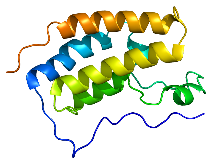
p53
BRD4
Presentation Overview
Thesis Defense | BRD4 & p53 in Breast Cancer
[University Name] | [Institute Name] | [Student Name] | [Enrollment ID] | [Department]
01
🔬
Introduction
Breast Cancer Overview, Epidemiology
02
🧬
Molecular Biology
BRD4, p53, BET Family
03
🔍
Epigenetics
Histone Modifications, Super-Enhancers
04
💊
MZ1 PROTAC
Mechanism & Selectivity
05
🏥
MCF-7 Model
Cell Line & DNA Damage
06
⚗️
Methodology
Western Blot, RT-PCR, rMATS
07
📊
Results
BCA, Western Blot, qPCR Data
08
💬
Discussion
Findings & Significance
09
🎯
Conclusion
Future Scope & References
CHAPTER 1
Introduction
2.3 Million
New cases annually (GLOBOCAN 2021)
685,000
Deaths per year worldwide
Most common
Cancer in women globally
TP53 Mutation
50% of all cancers
BRD4 Upregulation
Epigenetic driver
TNBC Subtype
Most aggressive, no receptor targets
Cell Cycle Dysregulation
CDK pathway disruption
The interplay between BRD4 and p53 represents a critical therapeutic axis in breast cancer progression.
[University Name] | [Student Name] | [Enrollment ID]
EPIDEMIOLOGY
Global & Indian Burden of Breast Cancer
2.3M
685K
192,020
98,337
Source: GLOBOCAN 2022
Luminal A
Luminal B
HER2-enriched
TNBC / Basal-like
[University Name] | [Institute Name] | [Department]
Molecular Classification
Four Subtypes — Distinct Biology, Distinct Outcomes
LUMINAL A
~40%
Low Ki-67
TP53 Mut: <15%
Endocrine therapy responsive
BEST PROGNOSIS
LUMINAL B
~20%
TP53 Mut: 25%
Endocrine + Chemo
MODERATE PROGNOSIS
HER2-ENRICHED
~15%
TP53 Mut: 40%
Trastuzumab
MODERATE PROGNOSIS
TNBC / BASAL-LIKE
~15-20%
TP53 Mut: >82%
Chemo only (PARP/PD1)
POOR PROGNOSIS
Department of Oncology | Graduate Research Defense
MOLECULAR BIOLOGY
BRD4 & the BET Protein Family
BRD2
BRD3
BRD4
OVEREXPRESSED IN CANCER
BRDT
UNIVERSITY OF CALIFORNIA
DEPARTMENT OF MOLECULAR BIOLOGY
12
BRD4 Dual Function Pathway
Transcription regulation and oncogene activation mechanics
TUMOR SUPPRESSOR
p53: The Guardian of the Genome
TP53 — Most Frequently Mutated Gene in Human Cancer
TP53 mutated in ~50% of all cancers
>80% of TNBC | <15% of Luminal A
University Name | Institute Name | Department of Molecular Biology
MOLECULAR AXIS
BRD4–p53 Regulatory Interaction
How BRD4 Controls the Tumor Suppressor
[Institute Logo]
[University Name] | [Institute Name] | [Student Name] | [Enrollment ID] | [Department]
BRD4 occupancy required for baseline TP53 transcription (Zhou et al., 2022)
BRD4 Active
BRD4 reads H3K27ac at TP53 super-enhancer
Recruits transcriptional machinery
p53 mRNA transcribed
p53 protein expressed
Tumor suppression active
BRD4 Inhibited (JQ1)
BRD4 displaced from super-enhancer
Partial reduction in TP53 transcription
Some p53 protein reduction
BRD4 Degraded (MZ1)
Complete BRD4 elimination
Super-enhancer collapse
Post-translational p53 accumulation
Complex regulatory outcome
Nagarajan et al. (2022)
BRD4 regulates wild-type p53 at super-enhancers in luminal breast cancer
Zhou et al. (2022)
BRD4 required for baseline TP53 transcription in MCF-7 cells
DNA DAMAGE RESPONSE
Radiation & DNA Double-Strand Break Repair
2 Gy Ionizing Radiation
DNA Double-Strand Break (DSB)
MRN Complex (MRE11-RAD50-NBS1) detects DSB
ATM Kinase Recruited & Activated
DNA Repair Pathway
γH2AX foci formation at DSBs
Homologous Recombination (HR) via RAD51
BRD4 phosphorylated at Ser1117 by IKKα → coordinates DDR
p53 Pathway
p53 phosphorylated at Ser15/Ser20
MDM2 interaction disrupted
p53 stabilized & accumulated
p21/CDK arrest → G1/S checkpoint
BAX/PUMA → Apoptosis
MCF-7 Response: G2/M arrest (not apoptosis) — Radioresistant apoptotic phenotype
BRD4 degradation by MZ1 → ↓DNMT1 + ↓RAD51 → Radiosensitization (Foran et al., 2025)
BRD4 needed for baseline p53 levels → determines radiation response
Molecular Pathways in Radiation Pathology | Breast Cancer Research
EPIGENETICS
Histone Modification: Writers, Erasers & Readers
The Epigenetic Code in Breast Cancer
Ac marks (acetylation)
Me marks (methylation)
Ph marks (phosphorylation)
Ub marks (ubiquitination)
WRITERS
Deposit activating/repressive marks
HATs
CBP/p300, GCN5, PCAF
HMTs
EZH2, MLL, G9a
H3K27ac ★, H3K4me3
ERASERS
Remove histone marks
HDACs
HDAC1/2, SIRT1
HDMs
LSD1/KDM1A
HDAC1 reduced by BRD4-RAC1 inhibition
READERS
Recognize & bind marked histones
BRD4 (BET proteins)
Reads H3K27ac marks
Chromodomains
Read methylation
PHD fingers
Read methylation marks
★ BRD4 is the key oncogenic reader → THERAPEUTIC TARGET
H3K27ac
BRD4 docking, active super-enhancers
H3K27me3
Repressive, antagonizes BRD4
H3K9ac
Active transcription
H3K9me2/3
DSB repair sites
H4K16ac
Reduced at radiation DSBs
University Name | Epigenetics & Breast Cancer Research | 2024
Super-Enhancers & BRD4 Overexpression in Breast Cancer
TCGA Analysis: BRD4 as an Epigenetic Oncogene
BRD4 mRNA Expression Across Breast Cancer Subtypes (TCGA)
Normal Mammary
1.0×
Luminal A
1.8×
Luminal B
2.4×
HER2+
3.1×
TNBC / Basal-like
4.6×
4.6× overexpression uniquely in TNBC
High BRD4 = High tumor grade + Lymph node infiltration + Reduced survival
Potential prognostic biomarker for optimal risk stratification
Compact Chromatin (Repressed)
Open Chromatin with Super-Enhancer
MYC
BCL2
CCND1
TP53
BET Inhibitors (JQ1, MZ1) → BRD4 removed from super-enhancer → Preferential repression of oncogenes
BRD4 upregulation drives endocrine therapy resistance in ER+ breast cancer via super-enhancer reprogramming of ESR1
UNIV LOGO
INST LOGO
[University Name] | [Institute Name] | Data source: TCGA Breast Cancer (BRCA) Project
[University Logo]
[Institute Logo]
EXPERIMENTAL MODEL
MCF-7: The Luminal A Breast Cancer Model
[University Name] | [Institute Name] | [Student Name] | [Enrollment ID] | [Department]
[UNIV]
[DEPT]
THERAPEUTIC TOOL
MZ1 — A PROTAC Degrader of BRD4
Induced Proximity → Ubiquitination → Proteasomal Degradation
[University Name] | [Department] | 2024
BRD4's post-translational regulation of p53 in ER+ breast cancer is poorly understood
No study examined BRD4 degradation (vs inhibition) effect on p53 in radiation context
rMATS alternative splicing analysis pipeline not established in lab for splicing modulator studies
To investigate the role of BRD4 in regulation of p53 expression in breast cancer cells (MCF-7) and to establish rMATS pipeline in lab to study splicing changes upon treatment with splicing modulators.
Evaluate effect of BRD4 degradation (MZ1) and ionizing radiation (2 Gy) on p53 PROTEIN expression in MCF-7 using Western blotting
Study effect of BRD4 degradation on p53 TRANSCRIPT expression using RT-PCR
Establish a functional rMATS pipeline for alternative splicing analysis
[University Name] | [Institute Name] | [Department]
Experimental Design
MCF-7 Cells | MZ1 PROTAC | Ionizing Radiation
MCF-7 (ER+, Wild-type p53) | DMEM + 10% FBS + 1% Pen/Strep | 37°C, 5% CO2 | 70-80% confluency
MZ1 Treatment
100 nM MZ1 (PROTAC BRD4 degrader)
Pre-treatment before radiation
Selective BRD4 degradation
8 Experimental Groups
Control
−MZ1 / −IR
MZ1 Only
+MZ1 / −IR
2Gy 30min
−MZ1 / +IR
MZ1+2Gy 30min
+MZ1 / +IR
2Gy 2hr
−MZ1 / +IR
MZ1+2Gy 2hr
+MZ1 / +IR
2Gy 4hr
−MZ1 / +IR
MZ1+2Gy 4hr
+MZ1 / +IR
Ionizing Radiation
2 Gy dose
Harvested at 3 time points: 30 min, 2 hr, 4 hr
Protein Analysis
Nuclear Protein Extraction
BCA Assay
Western Blot (p53 + α-tubulin)
Transcript Analysis
RNA Extraction
cDNA Synthesis
RT-PCR (p53/TBP)
Bioinformatics
rMATS Pipeline Setup
[University Logo]
[Institute Logo]
[University Name] | [Institute Name] | [Student Name] | [Enrollment ID] | [Department]
METHODOLOGY
Nuclear Protein Extraction & BCA Protein Assay
DEPARTMENT OF BIOCHEMISTRY | UNIVERSITY NAME
12
Western Blotting Protocol
SDS-PAGE → Transfer → Antibody Detection
Sample Preparation & SDS-PAGE
Denaturation at 95°C with 4× SDS loading dye + β-mercaptoethanol
12% resolving gel + 4% stacking gel
120V stacking → 100V resolving
Tris-glycine-SDS running buffer
PVDF Membrane Transfer
Wet transfer system
PVDF activated in methanol 15-20s
Transfer buffer: Methanol 20% + 1x Running buffer
Blocking
5% BSA in TBST — 1 hour RT
Prevents non-specific antibody binding
Antibody Incubation
Detection & Analysis
Li-Cor Odyssey Infrared Imaging System
Band densitometry: ImageJ software
Normalized to α-tubulin loading control
[University Name] | [Institute Name] | [Department]
MOLECULAR ANALYSIS
RNA Extraction & RT-PCR Protocol
RNA Extraction Flowchart (QIAGEN RNeasy Plus)
Cell Lysis
RLT buffer added to pellet, repeated pipetting, centrifuge 10,000 rpm/3 min
QIAshredder Column
Mechanical homogenization, reduce viscosity, remove debris
gDNA Eliminator Column
Removes genomic DNA contamination
Add 70% Ethanol
Promotes RNA binding to silica membrane
RNeasy Spin Column
RNA binds selectively, contaminants pass through
DNase Treatment
DNase + RDD buffer, 15 min RT, complete DNA digestion
Wash Steps
RW1 buffer washes, remove proteins
Elute with RNase-free water
Purified RNA collected
PURITY CHECK
Both samples: high purity, A260/280 ≈ 2.0 = RNA quality confirmed
cDNA SYNTHESIS
RT-PCR SETUP (SYBR Green)
PRIMERS
Analysis Method
[University Name] • [Institute Name] • Department of Biochemistry
rMATS Pipeline: Alternative Splicing Analysis
Offline Setup on WSL Ubuntu 20.04 | Differential Splicing Detection
Statistical framework for detecting differential alternative splicing from RNA-Seq data.
Compares splice junction reads between two conditions using replicate BAM files + GTF annotation.
Quality control of raw sequence data
Adapter removal & quality filtering
Splice-aware alignment to human genome
SAM → BAM conversion, sort & index
treated_bams.txt + control_bams.txt + GTF
5 distinct splicing event types
Ubuntu 20.04 (WSL)
Exon inclusion level difference
Statistical significance
Corrected for multiple testing
Pipeline Successfully Established
Framework ready for splicing modulator analysis
[University Name] | [Institute Name] | [Student Name] | Bioinformatics Core
RESULTS — SECTION 4.1 & 4.2
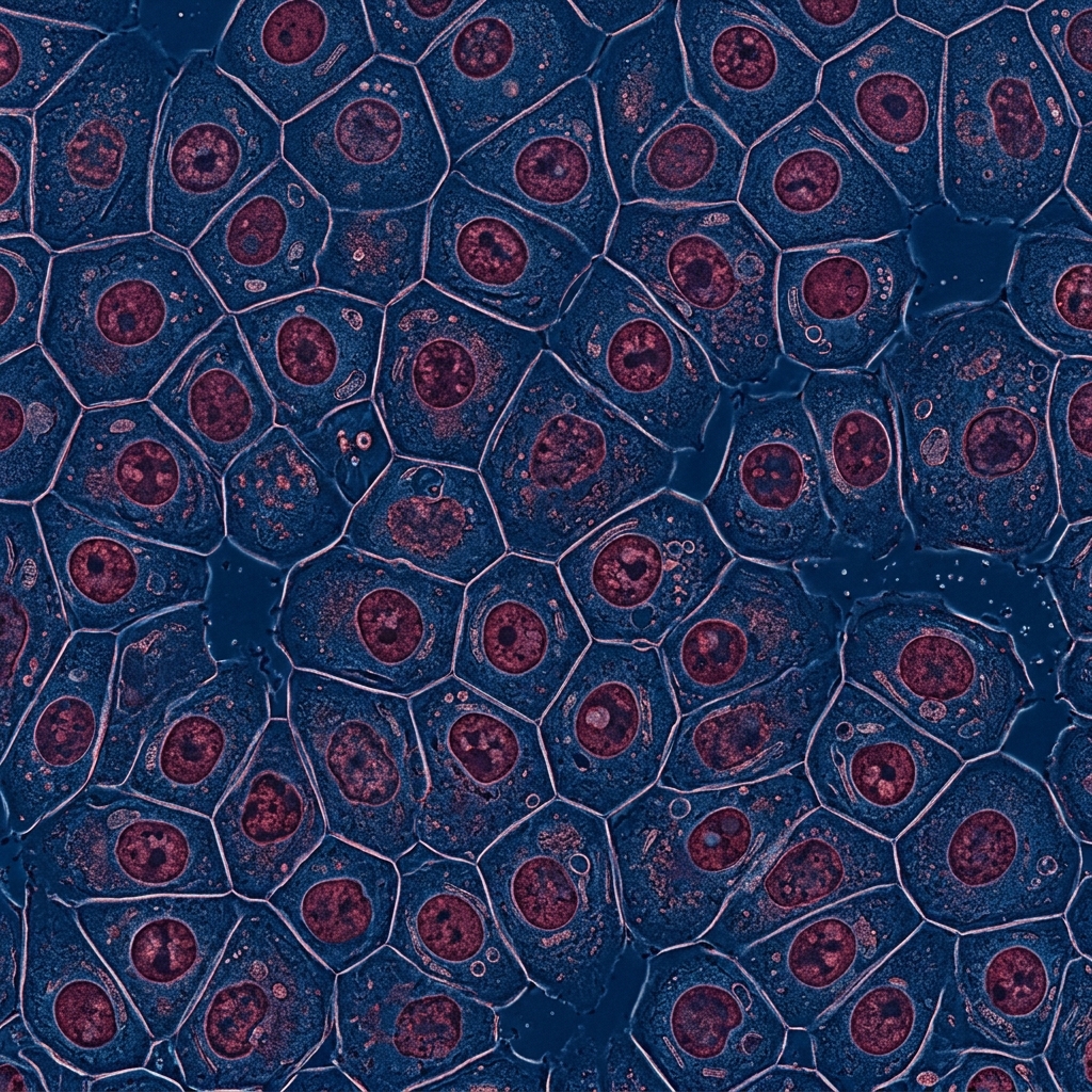
MCF-7 Cell Morphology & Protein Quantification
Adherent epithelial morphology confirmed
Polygonal to irregular shape — epithelial origin
Cobblestone arrangement — intact cell-cell adhesion
Active proliferation clusters visible
Cells suitable for downstream experiments
MZ1 + Radiation combinations show altered protein concentrations — treatment-dependent cellular responses confirmed
[University Name] | [Institute Name] | [Student Name] | [Enrollment ID] | [Department]
Western Blot Analysis: p53 Protein Expression
BRD4 Degradation (MZ1) Consistently Elevates p53 Levels
p53
BRD4
[University Name] | [Institute Name] | [Student Name] | [Enrollment ID] | [Department]
RESULTS — 4.4 & 4.5
RT-PCR: TP53 Transcript Analysis & Agarose Gel
[University Logo]
[Institute Logo]
RNA Quality Summary
1472.44 ng/µL | A260/280 = 2.094
1272.32 ng/µL | A260/280 = 2.103
p53
TBP
26.32
26.48
22.72
22.31
ΔCq = 3.60
ΔCq = 4.17
ΔΔCq = 0.56
Fold Change = 0.68
Fold change ≈ 0.68 — NOT significantly different from 1.0 (unity).
MZ1 does NOT alter TP53 mRNA levels.
No visible change in band intensity between control and MZ-1 samples → corroborates RT-PCR finding
MZ1 does NOT affect TP53 at transcriptional level → BRD4 regulates p53 POST-TRANSCRIPTIONALLY / POST-TRANSLATIONALLY (protein stability mechanism)
[University Name] | [Institute Name] | [Student Name] | [Enrollment ID] | [Department]
rMATS Pipeline: Alternative Splicing Output
Successfully Established Framework for Splicing Analysis
LOGO
LOGO
Bioinformatics Pipeline | Splicing Analysis | Transcriptomics
Application Context
Key Laboratory Milestone
This establishes the first rMATS workflow in the laboratory — enabling future transcriptome-wide splicing studies in cancer.
DISCUSSION
Interpretation of Findings
[University Logo]
[Institute Logo]
Key Finding 1: MZ1 Elevates p53 Protein
BRD4 degradation by MZ1 → Consistent p53 protein upregulation across all conditions
Strongest effect at 2hr post-2Gy radiation
Mechanism proposed: BRD4 regulates MDM2-p53 axis OR directly stabilizes p53 protein post-translationally
Supporting literature: Nagarajan et al. (2022), Zhou et al. (2022)
Key Finding 2: No Transcriptional Change
TP53 mRNA levels unchanged by MZ1 (fold change = 0.68, near unity)
BRD4 does NOT regulate p53 at transcriptional level in these conditions
Mechanistic implication: BRD4 may regulate p53 protein STABILITY via MDM2 degradation pathway or direct protein interaction
Paradigm: Protein-level ≠ mRNA-level regulation
MCF-7 Model Significance
Wild-type p53 — clean model for epigenetic regulation study
Luminal A → 40% of all breast cancers → high clinical relevance
Intact MDM2-p53 feedback loop allows clean interpretation
Results free from gain-of-function mutant p53 confound (unlike MDA-MB-231)
rMATS Pipeline Achievement
First rMATS workflow established in laboratory
Successfully processes RNA-Seq → BAM → Differential splicing events
Framework ready for: splicing modulator experiments, spliceome characterization
Foundation for future transcriptome-wide studies
Integrative Model: BRD4-p53 Regulation & Radiation Response
BRD4 Active
p53 Protein Regulated at Post-translational Level
Normal p53 Function
MZ1 Treatment
BRD4 Degraded
Altered p53 Protein Stability
Enhanced p53 Accumulation
Radiation
p53 Stabilized via ATM
Combined Effect Amplified with MZ1
[University Name] | [Institute Name] | [Student Name] | [Enrollment ID] | [Department]
BRD4 as a Post-Translational Regulator of p53 in Breast Cancer
Experimental model successfully established
MCF-7 cells cultured, nuclear protein isolation, BCA quantification, Western blotting, RNA extraction, RT-PCR all performed effectively — reliable data generated
BRD4 Degradation Increases p53 Protein
MZ1 treatment leads to increased p53 protein intensity in MCF-7 nuclear extracts → BRD4 plays a positive regulatory role in p53 protein expression
Enhanced effect observed under 2Gy radiation — peak at 2hr post-irradiation
BRD4 Does NOT Affect TP53 Transcription
qPCR fold change ≈ 0.68 (near unity) → MZ1 does not alter TP53 mRNA levels
BRD4 regulates p53 POST-TRANSCRIPTIONALLY or POST-TRANSLATIONALLY
rMATS Pipeline Successfully Established
First rMATS framework in the laboratory — bioinformatics infrastructure for future splicing studies validated
Therapeutic Implication
BRD4 may be a therapeutic target for enhancing radiosensitivity — particularly in tumors with aberrant p53 expression
Rationale for BRD4-targeted therapy combined with radiotherapy to overcome resistance
These findings highlight a previously underappreciated role of BRD4 in post-translational p53 regulation in hormone-responsive breast cancer
[University Logo]
[Institute Logo]
PhD Defense | Conclusion | BRD4 and p53 Regulation
FUTURE DIRECTIONS
Future Work & Research Scope
Mechanistic Validation
Co-immunoprecipitation (Co-IP) to confirm BRD4-MDM2 interaction
Ubiquitination assays — confirm BRD4 role in p53 proteasomal regulation
CHX chase experiment — p53 protein stability assay
Functional Assays
Apoptosis detection (Annexin V/PI)
Clonogenic survival assay post-MZ1+radiation
Cell cycle analysis (flow cytometry)
Validate protein changes → biological outcomes
ChIP-seq Analysis
ChIP-seq for BRD4 and H3K27ac at TP53 locus
Confirm BRD4 super-enhancer occupancy at TP53
Map epigenetic changes post-MZ1 treatment
Extension to TNBC
Study BRD4-p53 axis in MDA-MB-231 (mutant p53 R280K)
Compare regulation between wt-p53 (MCF-7) vs mutp53 (MDA-MB-231)
Determine subtype-specificity of BRD4-p53 regulation
In Vivo & Clinical Translation
MZ1 + tamoxifen combination in MCF-7 xenograft models
MZ1 + ionizing radiation in vivo — test radiosensitization
Clinical relevance: BRD4 as predictive biomarker for radiation response
Establishing BRD4 as a radiosensitization target could revolutionize treatment of ER+ breast cancer patients resistant to endocrine therapy
[ UNIVERSITY LOGO ]
[University Name] | [Institute Name] | [Student Name] | [Enrollment ID] | [Department]
Slide 24
References
Key Citations Supporting This Research
37 references
GLOBOCAN 2022 | WHO 2019 | WCRF 2025
[University Name] | [Institute Name] | [Student Name] | [Enrollment ID] | [Department]
1.
Ali et al. (2021) — Co-targeting BRD4 and RAC1.
Int J Biol Sci, 17(14), 3760–3779.
2.
Dillon et al. (2023) — BRD4 inhibition in ER+ breast cancer.
Cancers, 15(16), 4066.
3.
Frezzo et al. (2023) — BRD4-p53 signalling axis.
Int J Mol Sci, 24(7), 6201.
4.
Gadd et al. (2017) — PROTAC structural basis.
Nature Chem Biol, 13(5), 514–521.
5.
Kotekar & Bhatt (2023) — BRD4 and MYC.
FEBS J, 290(20), 4820–4842.
6.
Liu et al. (2022) — BRD4 post-translational modifications.
Front Oncol, 12, 847701.
7.
Ma et al. (2022) — MZ1 in AML.
Cancer Biol Ther, 23(1), 1–15.
8.
Nagarajan et al. (2022) — BRD4 and wt-p53 in luminal cancer.
Nucleic Acids Res, 50(11).
9.
Naeimzadeh et al. (2024) — Mutant p53 in TNBC.
Cell Commun Signal, 22, 484.
10.
Qian et al. (2023) — Super-enhancers and BRD4.
Cell Death Discov, 9(1), 470.
11.
Wan et al. (2022) — BRD4 super-enhancer in breast cancer.
PNAS, 119(6).
12.
Wang et al. (2023) — BET proteins: biology and therapy.
Signal Transduct, 8, 420.
13.
Yousuf & Khan (2025) — MDM2-p53 in breast cancer.
Oncology Res, 33(4).
14.
Zengerle et al. (2015) — MZ1 selective BRD4 degradation.
ACS Chem Biol, 10(8).
15.
Zhou et al. (2022) — BRD4 and mutant p53 in TNBC.
Int J Mol Sci, 23(23).
16.
Arnold et al. (2022) — Global breast cancer burden.
The Breast, 66, 15–23.
17.
Essmann et al. (2004) — MCF-7 radiation apoptosis resistance.
Cancer Res, 64(19).
18.
Floyd et al. (2013) — BRD4 insulates chromatin from DDR.
Nature, 498(7453).
[Univ<br>Logo]
[Inst<br>Logo]
Thank You
Questions & Discussion
Research Summary
🔬
BRD4 positively regulates p53 protein in MCF-7 cells
🧬
Regulation is post-translational, not transcriptional
⚗️
rMATS pipeline successfully established
[Student Name]
[student.email@university.edu]
[Department of Biochemistry / Biotechnology]
[Institute Name | University Name]
[Dr. Guide Name]
[Department | Institute | Year]
[University Name] | [Institute Name] | [Enrollment ID] | [Department]
- breast-cancer-research
- p53-protein
- brd4-inhibitor
- mcf-7-cells
- oncology
- epigenetics
- molecular-biology
- biomedical-science