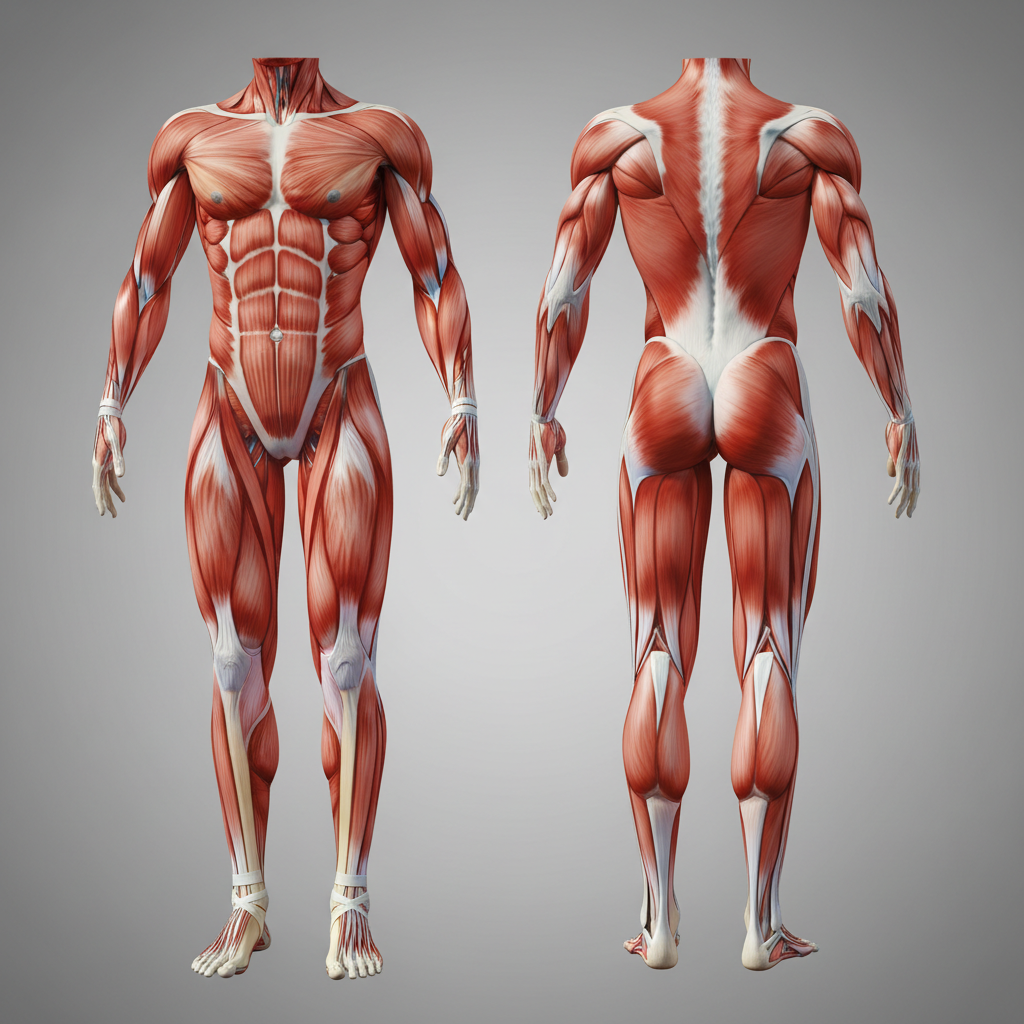
Lower Limb Anatomy: Muscles, Origins, and Functions
A comprehensive guide to lower limb muscles, covering the hip, thigh, leg, and foot with detailed origin and insertion points for students and clinicians.
Comprehensive Breakdown of Lower Limb Muscles
An Overview of Anatomy and Function
Presentation Outline
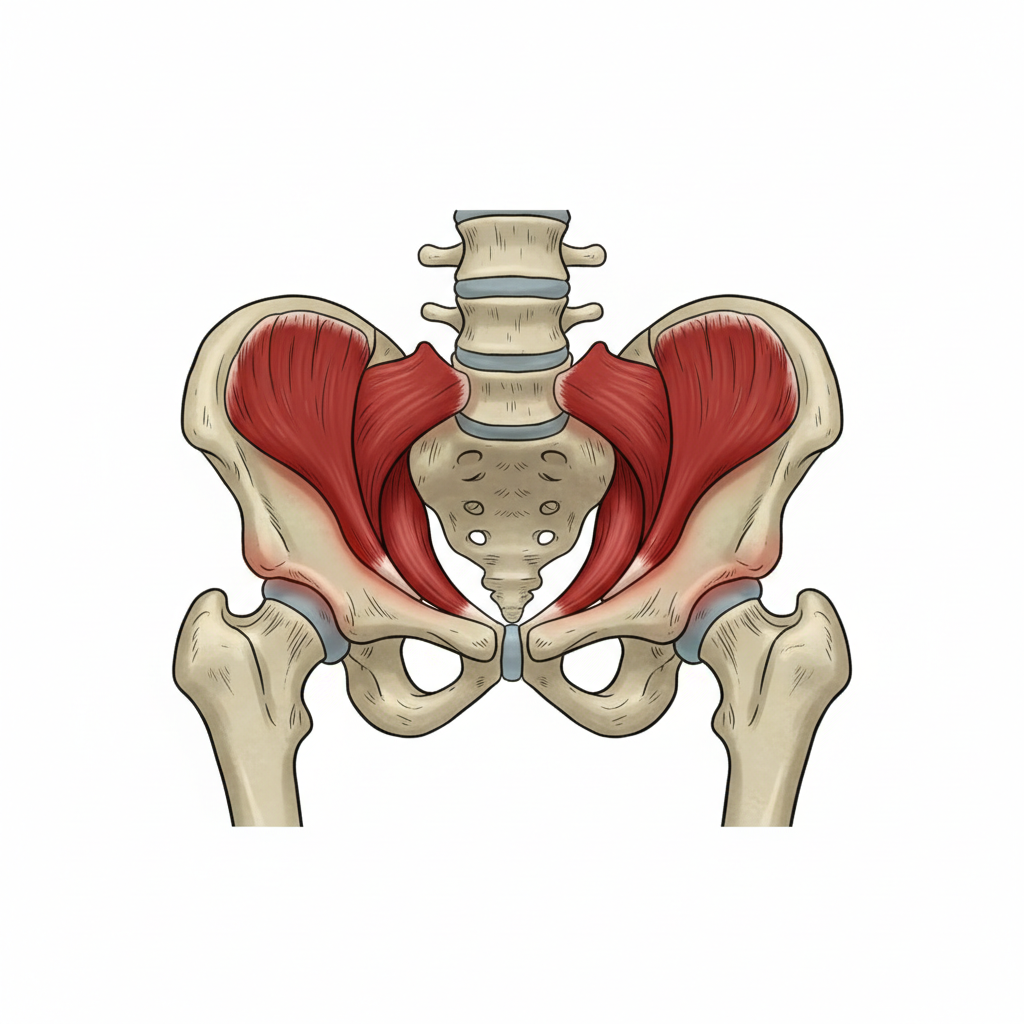
1. Hip Muscles
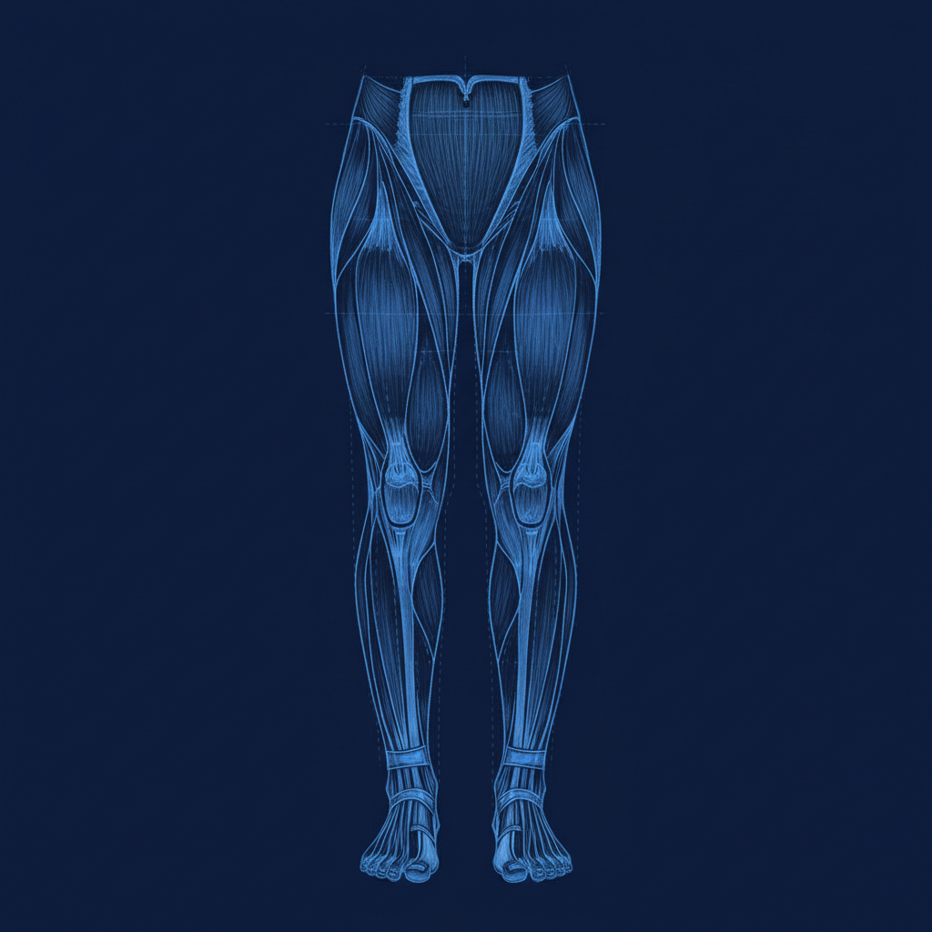
2. Thigh Muscles
3. Leg Muscles
4. Foot Muscles
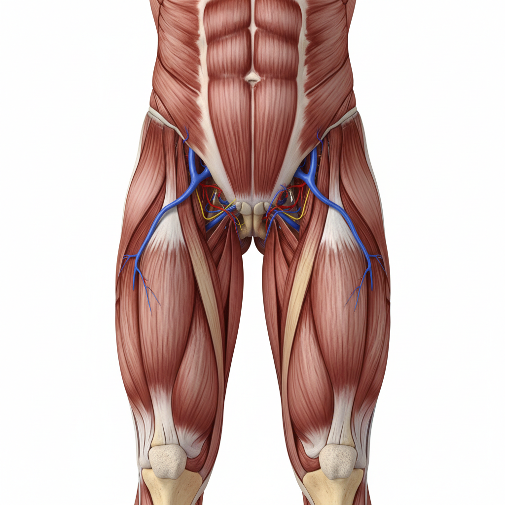
Hip Muscles: Flexors
Iliopsoas
<b>Origin:</b> Iliac fossa and lumbar vertebrae<br><b>Insertion:</b> Lesser trochanter of the femur
Rectus Femoris
<b>Origin:</b> Anterior inferior iliac spine (AIIS)<br><b>Insertion:</b> Tibial tuberosity via the patellar tendon
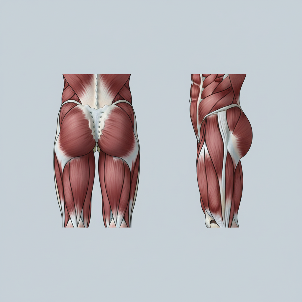
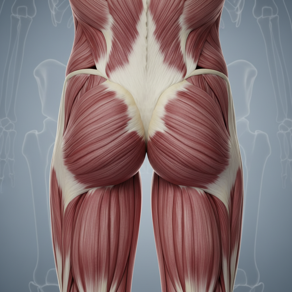
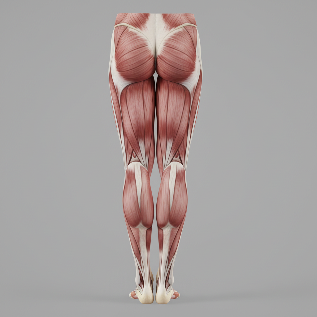
Hip Muscles: Extensors & Abductors
Hip Extensors
<b>Gluteus Maximus:</b> Origin from posterior iliac crest, sacrum, coccyx. Inserts at gluteal tuberosity and iliotibial tract.
Hip Abductors
<b>Gluteus Medius:</b> Origin from iliac crest/outer ilium. Inserts at greater trochanter.
<b>Gluteus Minimus:</b> Origin from outer ilium. Inserts at greater trochanter.
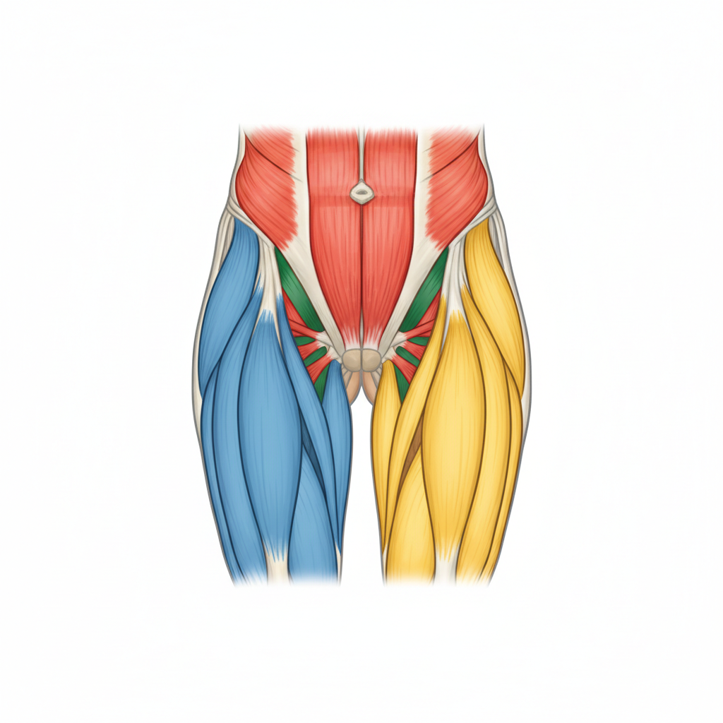
Hip Muscles: The Adductors
<strong style='color:#0052cc; font-size:32px;'>Adductor Magnus</strong><br>Origin: Ischium and pubis<br>Insertion: Linea aspera of the femur
<strong style='color:#0052cc; font-size:32px;'>Adductor Longus</strong><br>Origin: Pubis<br>Insertion: Middle third of the linea aspera
<strong style='color:#0052cc; font-size:32px;'>Adductor Brevis</strong><br>Origin: Pubis<br>Insertion: Superior part of the linea aspera
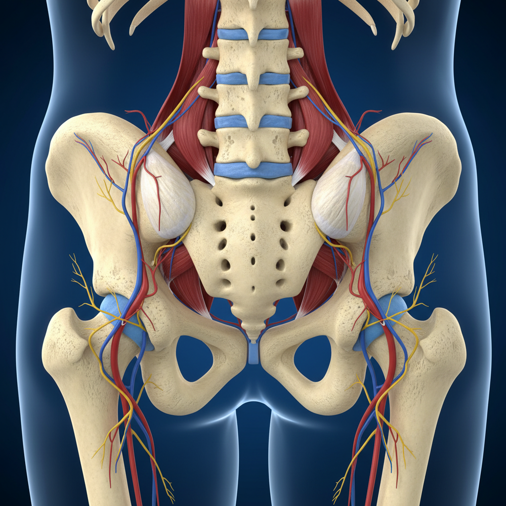
Hip Muscles: Rotators
<div style='margin-bottom:20px;'><strong style='color:#0052cc; display:block; margin-bottom:5px;'>Piriformis</strong>Origin: Anterior sacrum / Insertion: Greater trochanter</div><div style='margin-bottom:20px;'><strong style='color:#0052cc; display:block; margin-bottom:5px;'>Obturator Internus</strong>Origin: Inner obturator membrane / Insertion: Greater trochanter</div><div style='margin-bottom:20px;'><strong style='color:#0052cc; display:block; margin-bottom:5px;'>Gemellus Superior & Inferior</strong>Origin: Ischial spine/tuberosity / Insertion: Greater trochanter</div><div><strong style='color:#0052cc; display:block; margin-bottom:5px;'>Quadratus Femoris</strong>Origin: Ischial tuberosity / Insertion: Intertrochanteric crest</div>
Thigh Muscles: Quadriceps
The Knee Extensors
<b>Rectus Femoris</b><br>Origin: AIIS<br>Insertion: Tibial Tuberosity
<b>Vastus Lateralis</b><br>Origin: Greater Trochanter<br>Insertion: Tibial Tuberosity
<b>Vastus Medialis</b><br>Origin: Medial Linea Aspera<br>Insertion: Tibial Tuberosity
<b>Vastus Intermedius</b><br>Origin: Femoral Shaft<br>Insertion: Tibial Tuberosity
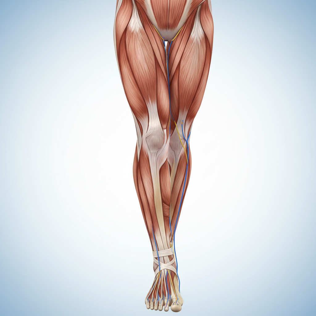
Thigh Muscles: Hamstrings
The Knee Flexors
Biceps Femoris
Origin: Ischial tuberosity & Linea aspera Insertion: Head of the fibula
Semitendinosus
Origin: Ischial tuberosity Insertion: Proximal tibia (pes anserinus)
Semimembranosus
Origin: Ischial tuberosity Insertion: Medial condyle of the tibia
Leg Muscles: Anterior Compartment
Dorsiflexors
<div style='background:white; padding:25px; margin-bottom:20px; border-radius:8px;'><h3 style='margin:0 0 10px 0; color:#0052cc;'>Tibialis Anterior</h3><p style='margin:0; color:#555;'>Lateral condyle/tibia → Medial cuneiform/1st metatarsal</p></div><div style='background:white; padding:25px; margin-bottom:20px; border-radius:8px;'><h3 style='margin:0 0 10px 0; color:#0052cc;'>Extensor Hallucis Longus</h3><p style='margin:0; color:#555;'>Middle fibula → Big toe distal phalanx</p></div><div style='background:white; padding:25px; border-radius:8px;'><h3 style='margin:0 0 10px 0; color:#0052cc;'>Extensor Digitorum Longus</h3><p style='margin:0; color:#555;'>Lat. condyle/fibula → Toes 2-5 phalanges</p></div>
Leg Muscles: Lateral & Posterior
Lateral (Evertors)
• Fibularis Longus<br>• Fibularis Brevis
Posterior (Plantar Flexors)
• Gastrocnemius (Two heads)<br>• Soleus (Deep to gastrocnemius)<br>• Tibialis Posterior (Deepest)
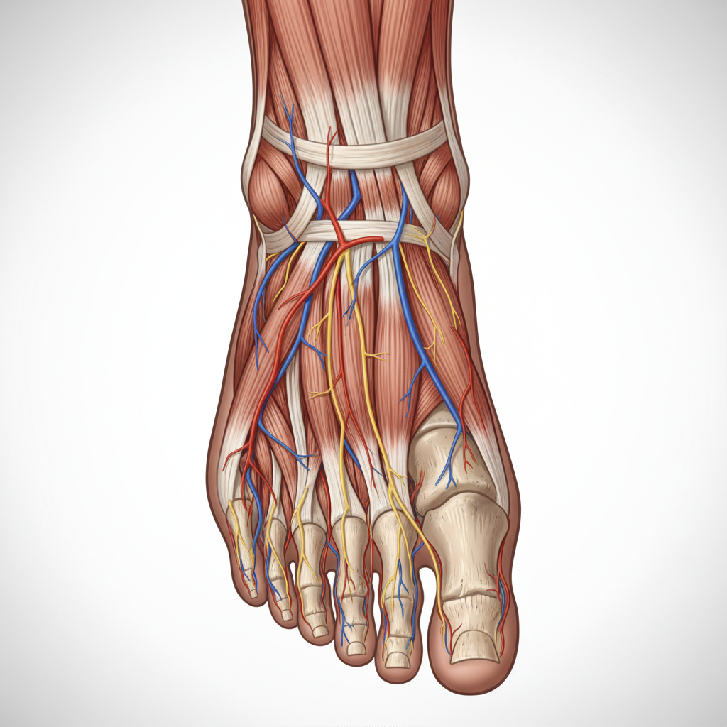
Foot Muscles: Intrinsic Group
Flexor Hallucis Brevis
Cuboid/cuneiforms → Big toe proximal phalanx
Flexor Digitorum Brevis
Calcaneus → Middle phalanges 2-5
Abductor Hallucis
Calcaneus → Medial base of big toe
Conclusion
The lower limb requires a complex interplay of hip, thigh, leg, and foot muscles to maintain stability and facilitate locomotion. Understanding these origins, insertions, and compartments is crucial for clinical diagnosis and rehabilitation.
- anatomy
- lower-limb
- muscle-physiology
- hip-muscles
- quadriceps
- hamstrings
- medical-education
- kinesiology