

Understanding Methemoglobinemia: Causes and Treatments
Learn about methemoglobinemia pathophysiology, diagnosis via the saturation gap, and treatments like methylene blue in this medical overview.
Methemoglobinemia
Pathophysiology, Clinical Presentation, and Management
Definition & Overview
Methemoglobinemia is a blood disorder in which an abnormal amount of methemoglobin is produced. Hemoglobin is the protein in red blood cells that carries and distributes oxygen to the body. <br><br>In this condition, the hemoglobin can carry oxygen but is <b>unable to release it effectively</b> to body tissues.
Pathophysiology: The Iron State
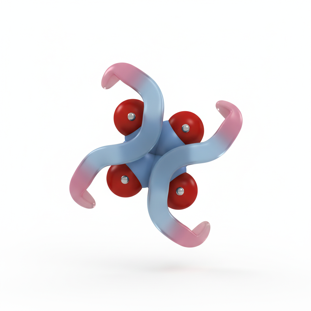
Normal Hemoglobin contains iron in the reduced <b>Ferrous (Fe2+)</b> state.
Methemoglobin contains iron in the oxidized <b>Ferric (Fe3+)</b> state.
Fe3+ creates a tight bind with oxygen, shifting the dissociation curve to the left.
Result: Tissue hypoxia despite adequate blood oxygen saturation.
Acquired Causes
Acquired Methemoglobinemia is far more common than congenital forms. It is typically caused by exposure to oxidizing agents:<br><br><ul><li><b>Medications:</b> Benzocaine (local anesthetic sprays), Dapsone, Sulfonamides, Nitroglycerin.</li><li><b>Environmental:</b> Nitrates (often found in contaminated well water), Anilin dyes, Naphthalene (mothballs).</li></ul>
Clinical Correlation: MetHb Level vs Symptoms
Clinical presentation correlates directly with the percentage of Methemoglobin. At low levels (<15%), patients may be asymptomatic but appear cyanotic. As levels rise, tissue hypoxia causes dyspnea, confusion, seizures, and eventually death.
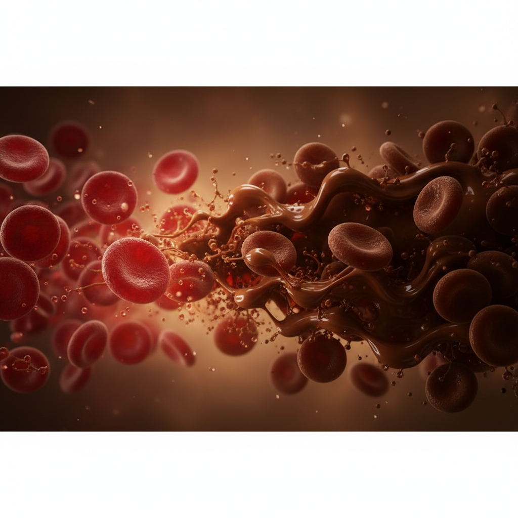
"Chocolate Cyanosis"
Classic Clinical Sign
Blood drawn from a patient with significant methemoglobinemia appears chocolate-brown and does not redden upon exposure to room air.
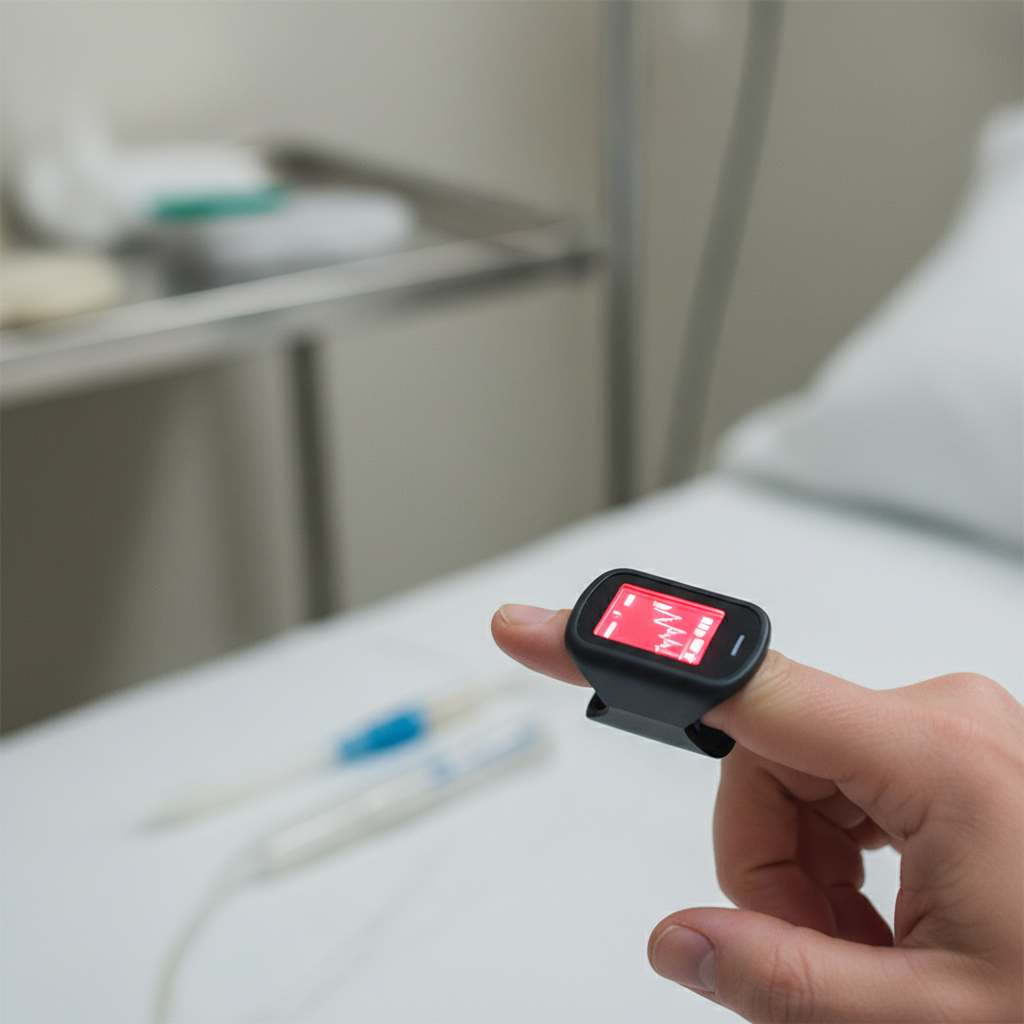
Diagnosis: The Saturation Gap
Standard Pulse Oximetry (SpO2) is unreliable. It only measures light absorption at two wavelengths (660nm and 940nm). Methemoglobin absorbs light equally at both.<br><br><b>The Result:</b> SpO2 falsely plateaus around 85%, regardless of the true oxygen saturation.<br><br><b>The Solution:</b> Co-oximetry is required to distinguish methemoglobin from oxyhemoglobin.
Primary Treatment: Methylene Blue
<b>Dosage:</b> 1-2 mg/kg IV over 5 minutes.<br><br><b>Mechanism of action:</b> Methylene blue acts as a cofactor for the enzyme NADPH-MetHb reductase. It uses NADPH to reduce itself to leukomethylene blue, which in turn reduces methemoglobin (Fe3+) back to hemoglobin (Fe2+).<br><br><b>Response:</b> Typically rapid response within 30-60 minutes.
Contraindications & Alternatives
<b>G6PD Deficiency:</b> Methylene blue is ineffective and dangerous; it requires NADPH (produced by G6PD) and can induce severe hemolysis.
<b>Alternative: Vitamin C (Ascorbic Acid):</b> Used if Methylene Blue is contraindicated. Dose: 1.5-2g IV. Note: Much slower action.
<b>Exchange Transfusion:</b> Reserved for severe unresponsive cases or massive hemolysis.
<b>Hyperbaric Oxygen:</b> Increases dissolved oxygen in plasma to sustain life significantly.
Summary & Prognosis
Methemoglobinemia is a potentially life-threatening but treatable condition. Recognition of the "Saturation Gap" (Low SpO2 on Pulse Ox vs Normal PaO2 on ABG) is the key diagnostic clue.<br><br>With prompt administration of Methylene Blue and removal of the offending agent, the prognosis is excellent for most acquired cases.
- methemoglobinemia
- pathophysiology
- methylene-blue
- hematology
- medical-education
- cyanosis
- blood-disorder