


The Lymphatic System: Anatomy, Physiology, and Immunity
Explore the lymphatic system’s role in fluid balance, immunity, and lipid transport. Covers major organs like the thymus, spleen, and lymph nodes.
The Lymphatic System
Anatomy, Physiology, and Immunity
Learning Objectives
Identify major lymphatic organs and vessels.
Understand the mechanism of lymph formation and flow.
Explain the role of the system in immunity and lipid transport.
What is the Lymphatic System?
A network of vessels, tissues, and organs that facilitates fluid return to the blood, immune defense, and lipid absorption.
Daily Fluid Balance
Of the 20L of fluid filtered from blood capillaries daily, the lymphatic system returns the 3L that is not strictly reabsorbed by veins.
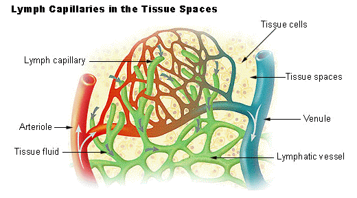
Lymphatic Capillaries
These blind-ended tubes are located in spaces between cells. They are more permeable than blood capillaries, allowing proteins and cell debris to enter. Key Feature: Endothelial cells overlap to form minivalves.
Mechanism of Entry: Anchoring Filaments
1. Interstitial fluid pressure rises. 2. Anchoring filaments pull endothelial flaps open. 3. Fluid enters the capillary (now called 'Lymph'). 4. Internal pressure closes the flaps, preventing backflow.
Lymphatic Trunks
• Lumbar Trunks (Lower limbs, pelvis) • Intestinal Trunk (Gastrointestinal organs) • Bronchomediastinal Trunks (Thorax) • Subclavian Trunks (Upper limbs) • Jugular Trunks (Head and neck)
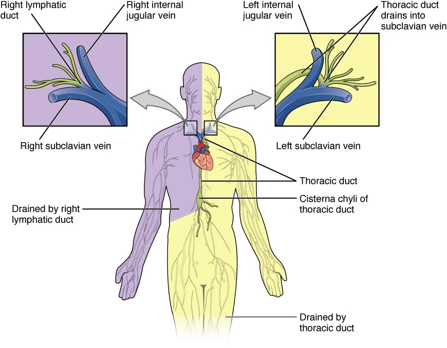
The Two Collecting Ducts
Right Lymphatic Duct
Drains lymph from the right upper quadrant ONLY: - Right side of the head and neck - Right upper limb - Right side of the thorax
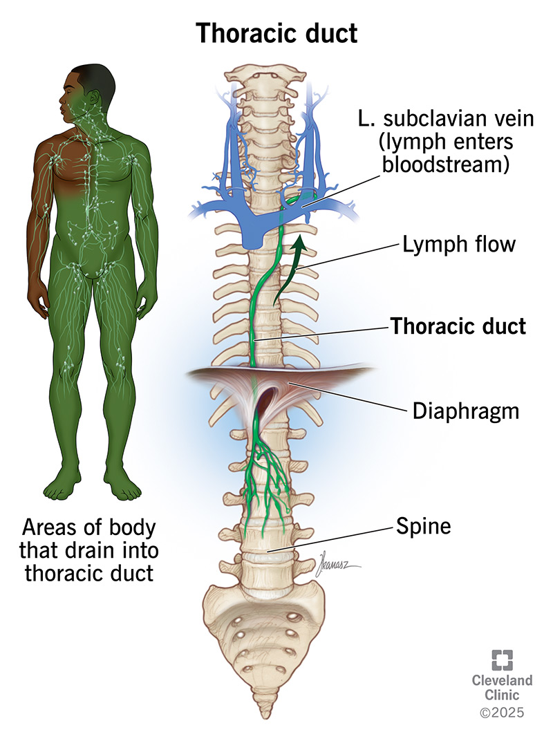
Thoracic Duct
Drains the REST of the body (approx. 75% of lymph). - Begins at the Cisterna Chyli (L1-L2 vertebra). - Ascends along the vertebral bodies. - Empties into the junction of the left internal jugular and left subclavian veins.
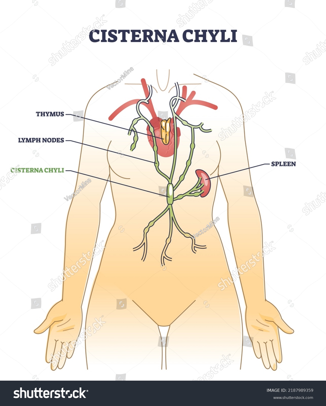
Cisterna Chyli
An enlarged sac at the base of the thoracic duct. It collects lymph from the two lumbar trunks (lower limbs) and the intestinal trunk (fat-rich chyle).
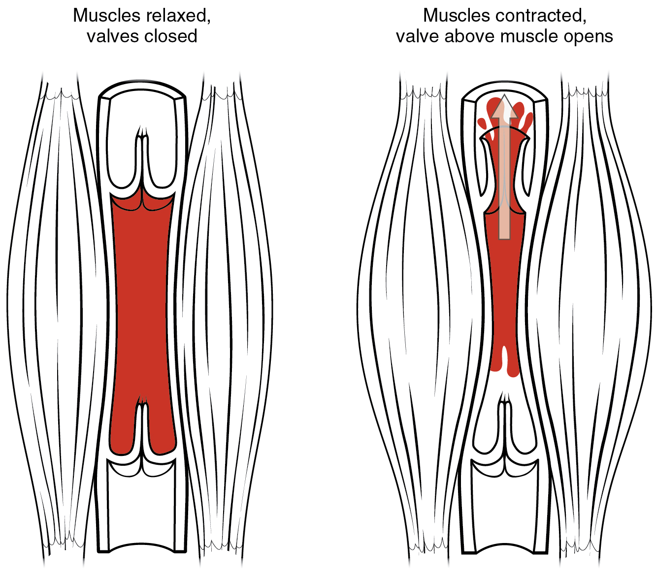
Factors Promoting Lymph Flow
Since the lymphatic system lacks a heart pump, it relies on: 1. Skeletal Muscle Pump: Muscles 'milk' the vessels. 2. Respiratory Pump: Pressure changes during breathing. 3. Valves: Prevent backflow. 4. Pulsation of adjacent arteries.
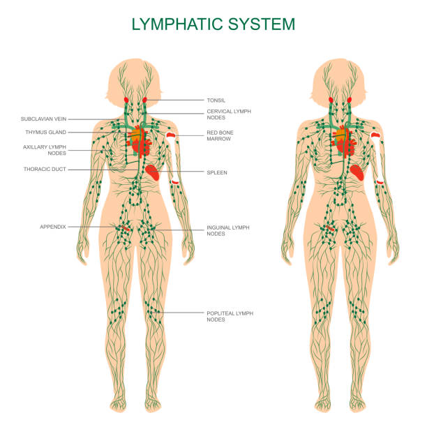
Lymphoid Organs Overview
PRIMARY (Development)
- Bone Marrow
- Thymus
SECONDARY (Activation)
- Lymph Nodes
- Spleen
- MALT (Tonsils, Peyer's Patches)
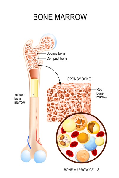
Red Bone Marrow
Site of Hematopoiesis (blood cell formation). Key Function: - Origin of ALL lymphocytes. - Site where B Cells mature (B for Bone).
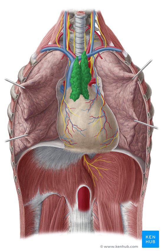
The Thymus Gland
Located in the mediastinum, above the heart.
Site where T Cells mature and learn to distinguish 'self' from 'non-self'.
Large in infants, it atrophies (shrinks) significantly in adults.
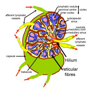
Lymph Nodes: The Filters
Cluster Areas: - Cervical (Neck) - Axillary (Armpit) - Inguinal (Groin)
Lymph Node Anatomy
• Afferent Vessels (Many in) • Efferent Vessels (Few out - slows flow) • Cortex (B Cells) • Paracortex (T Cells) • Medulla
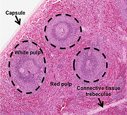
The Spleen
Largest lymphoid organ. Located in left upper quadrant. - White Pulp: Immune function (lymphocytes on reticular fibers). - Red Pulp: Filters old RBCs and platelets; stores iron.
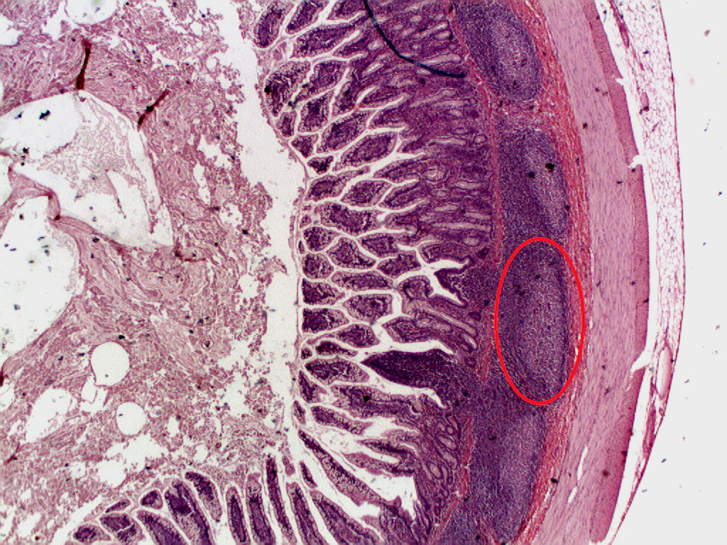
MALT: Mucosa-Associated Lymphoid Tissue
Lymphoid tissues located in mucous membranes to protect open passages. Examples: 1. Tonsils (Pharynx) 2. Peyer's Patches (Small Intestine) 3. Appendix
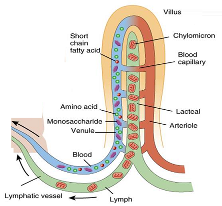
Physiology: Lipid Absorption
Most nutrients enter blood capillaries directly. However, large fats (lipids) cannot. Lacteals: Specialized lymphatic capillaries in intestinal villi absorb fats.
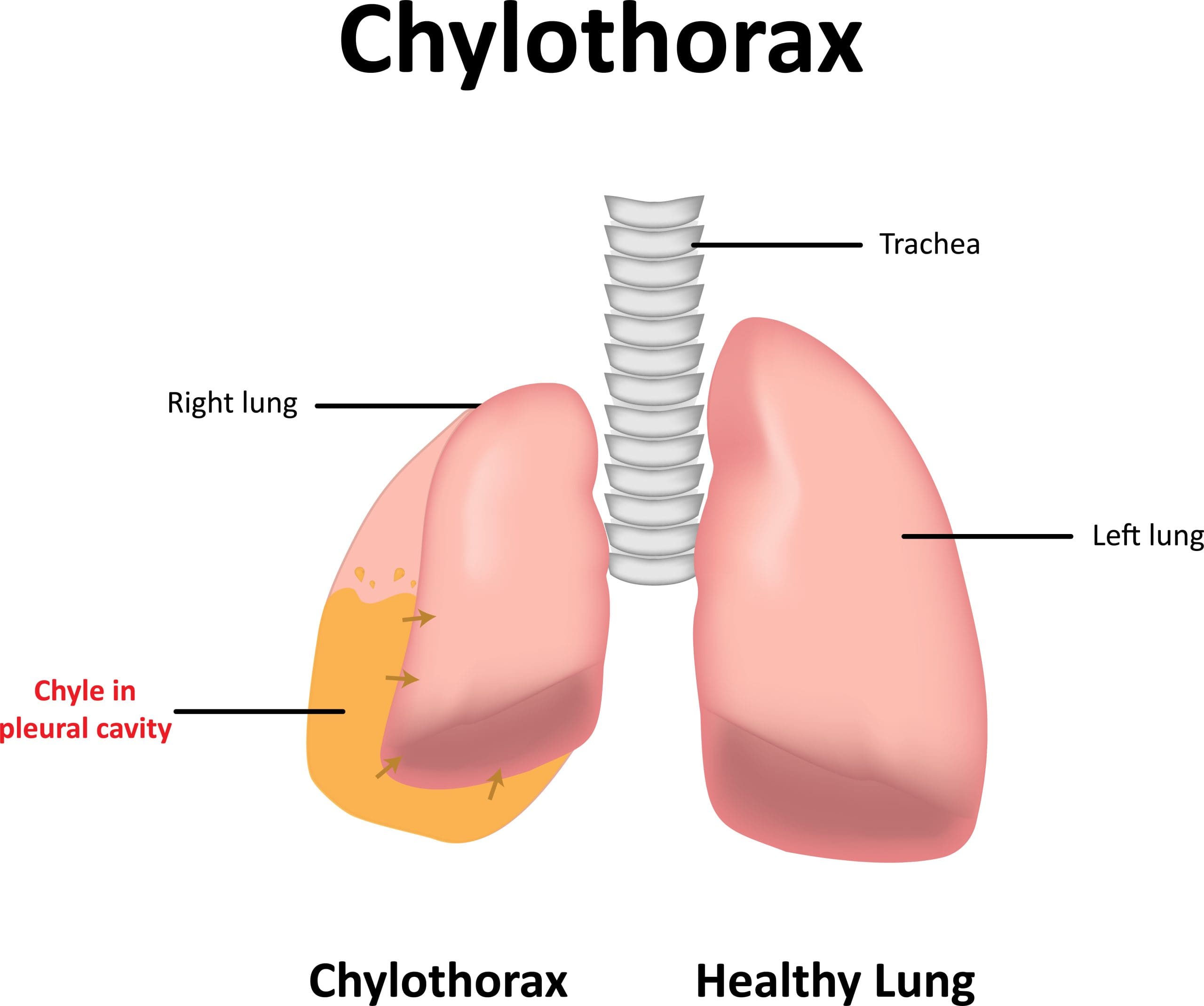
What is Chyle?
Chyle is lymph that is milky white because it is rich in lipids (chylomicrons). Pathway: Lacteals -> Intestinal Trunk -> Cisterna Chyli -> Thoracic Duct -> Blood Stream.
Immune Cells
- T Lymphocytes: Manage immune response, attack infected cells. - B Lymphocytes: Produce plasma cells which secrete antibodies. - Macrophages: Phagocytose (cell-eat) foreign substances.
Lymphocyte Distribution
T-Cells are the most abundant lymphocytes circulating in the blood.
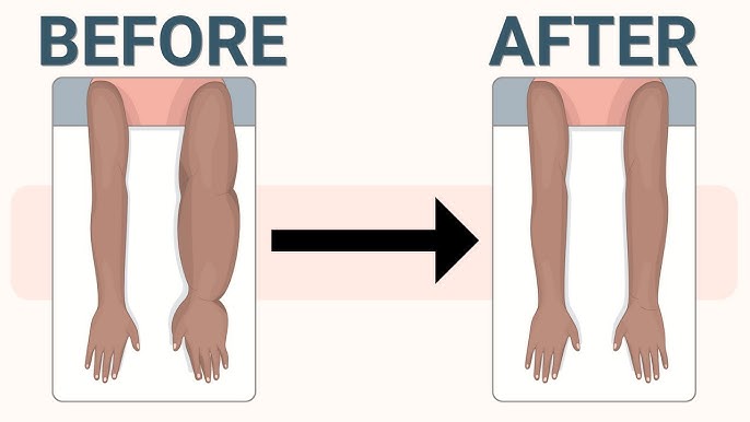
Clinical Insight: Lymphedema
Severe localized edema causing swelling. Causes: - Tumor blockage - Surgical removal of lymph nodes (e.g., in breast cancer) - Parasitic infection (Filariasis)
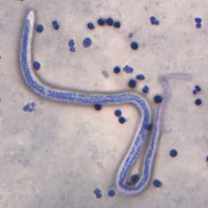
Elephantiasis (Wuchereria bancrofti)
A tropical parasitic disease transmitted by mosquitoes. The filarial worms block lymphatic vessels, causing extreme hypertrophy of the skin and subcutaneous tissues.
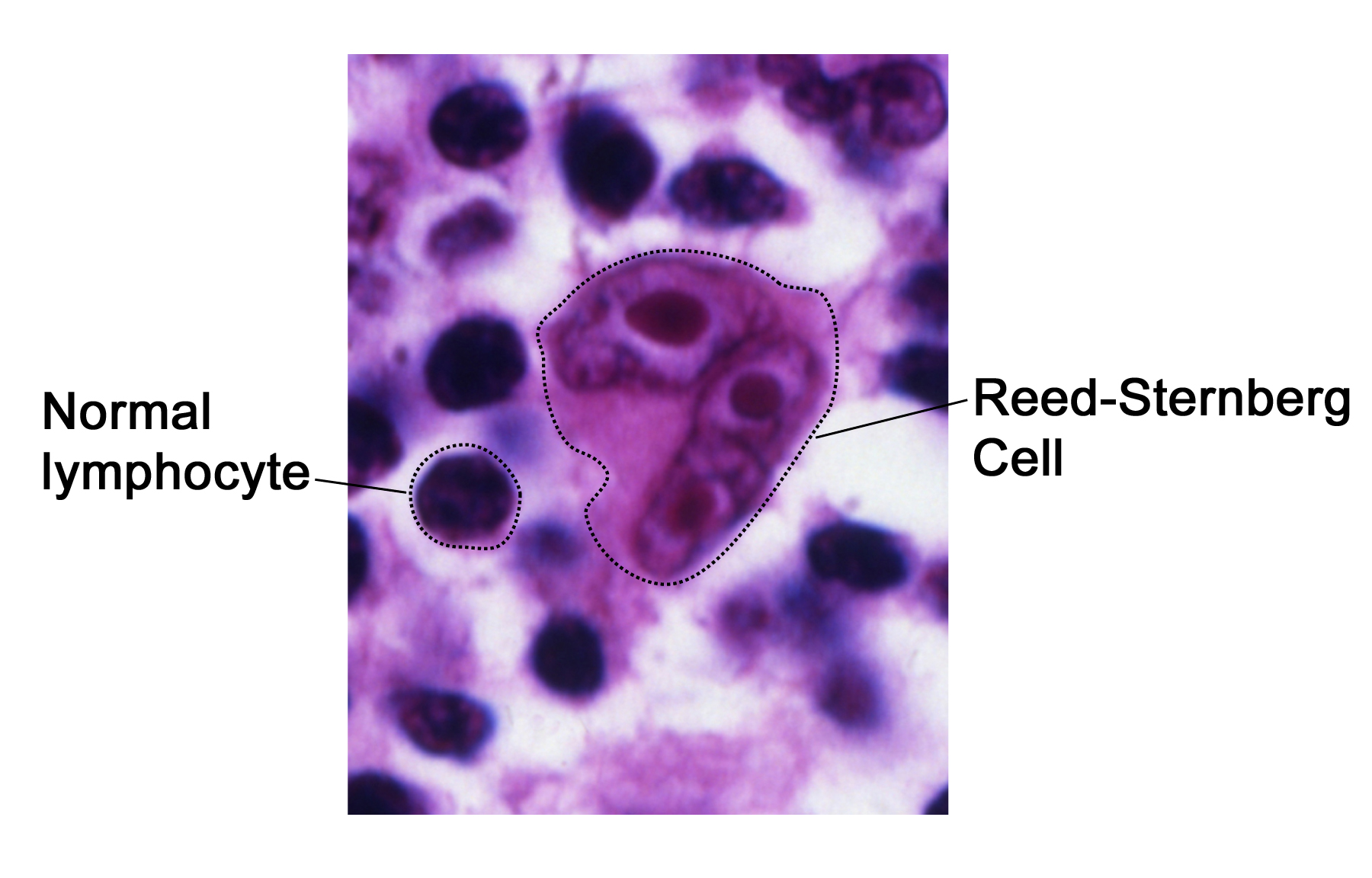
Clinical Insight: Lymphoma
Cancer of the lymph nodes. 1. Hodgkin's Lymphoma: Giant 'Reed-Sternberg' cells present. Often curable. 2. Non-Hodgkin's Lymphoma: More common, involves uncontrolled multiplication of lymphocytes.
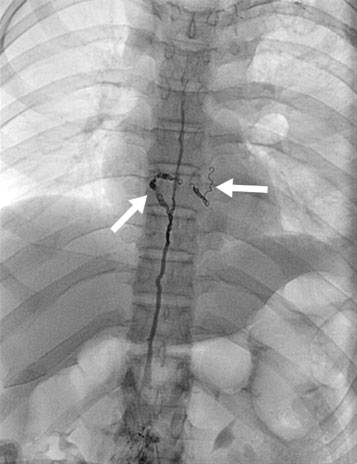
Diagnostic Imaging
Lymphangiography: An X-ray examination of the lymphatic vessels and nodes after injection of a contrast medium (dye). Useful for staging lymphomas.
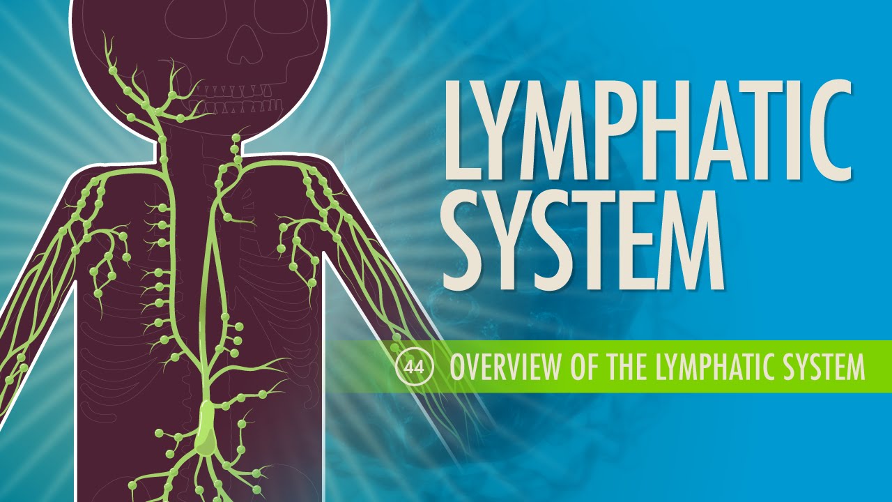
Video Overview
Recommended Viewing: Crash Course A&P: The Lymphatic System
https://www.youtube.com/watch?v=I7orwMgTQ5I
Summary
1. Fluid Return: Returns leaked 3L of plasma to blood. 2. Immunity: Nodes filter lymph; Spleen filters blood. 3. Lipid Absorption: Lacteals absorb dietary fats. 4. One-Way Flow: Capillaries -> Trunks -> Ducts -> Veins.
References & Further Study
• NIH (National Institutes of Health) • OpenStax Anatomy & Physiology • Kenhub: Lymphatic System Anatomy • Crash Course A&P (YouTube)
- anatomy
- physiology
- lymphatic-system
- immunity
- biology-education
- medical-science
- lymph-nodes