
Lymphatic System Anatomy and Physiology: A Comprehensive Guide
Explore the lymphatic system's anatomy, from fluid dynamics and ducts to lymph nodes, primary organs like the spleen, and its role in immunity and drainage.
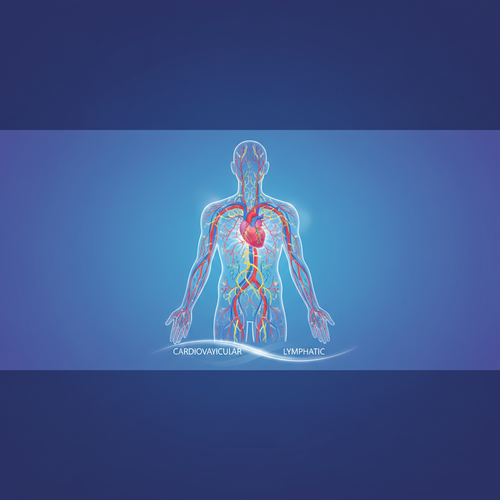
The Lymphatic System
Anatomy, Circulation, and Physiological Function
Human Anatomy & Physiology 101
Primary Functions
Fluid Balance: Returns excess interstitial fluid to the bloodstream.
Immunity: Houses lymphocytes for body defense against pathogens.
Lipid Absorption: Lacteals in the small intestine absorb dietary fats.
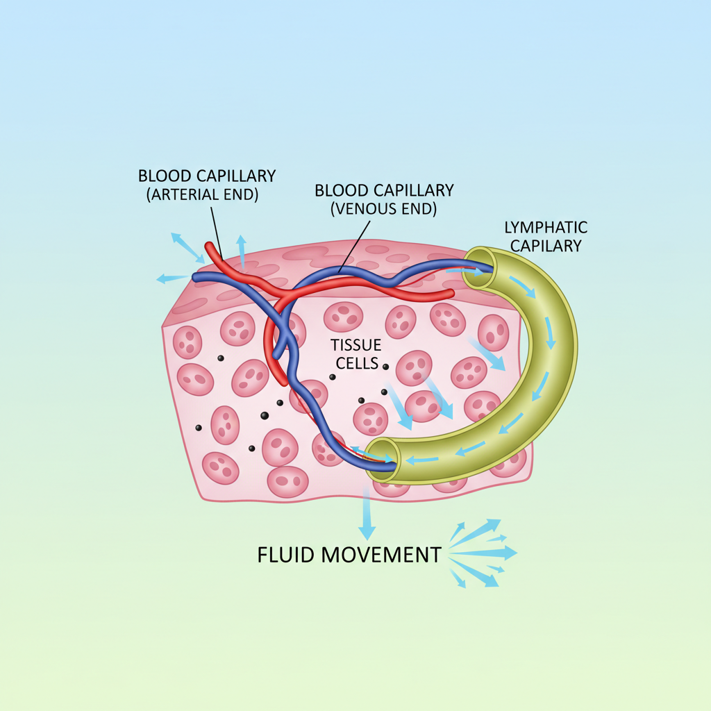
Daily Fluid Dynamics
Approximately 20 liters of plasma is filtered daily. The venous system reabsorbs about 85%, leaving 3 liters for the lymphatic system to return.
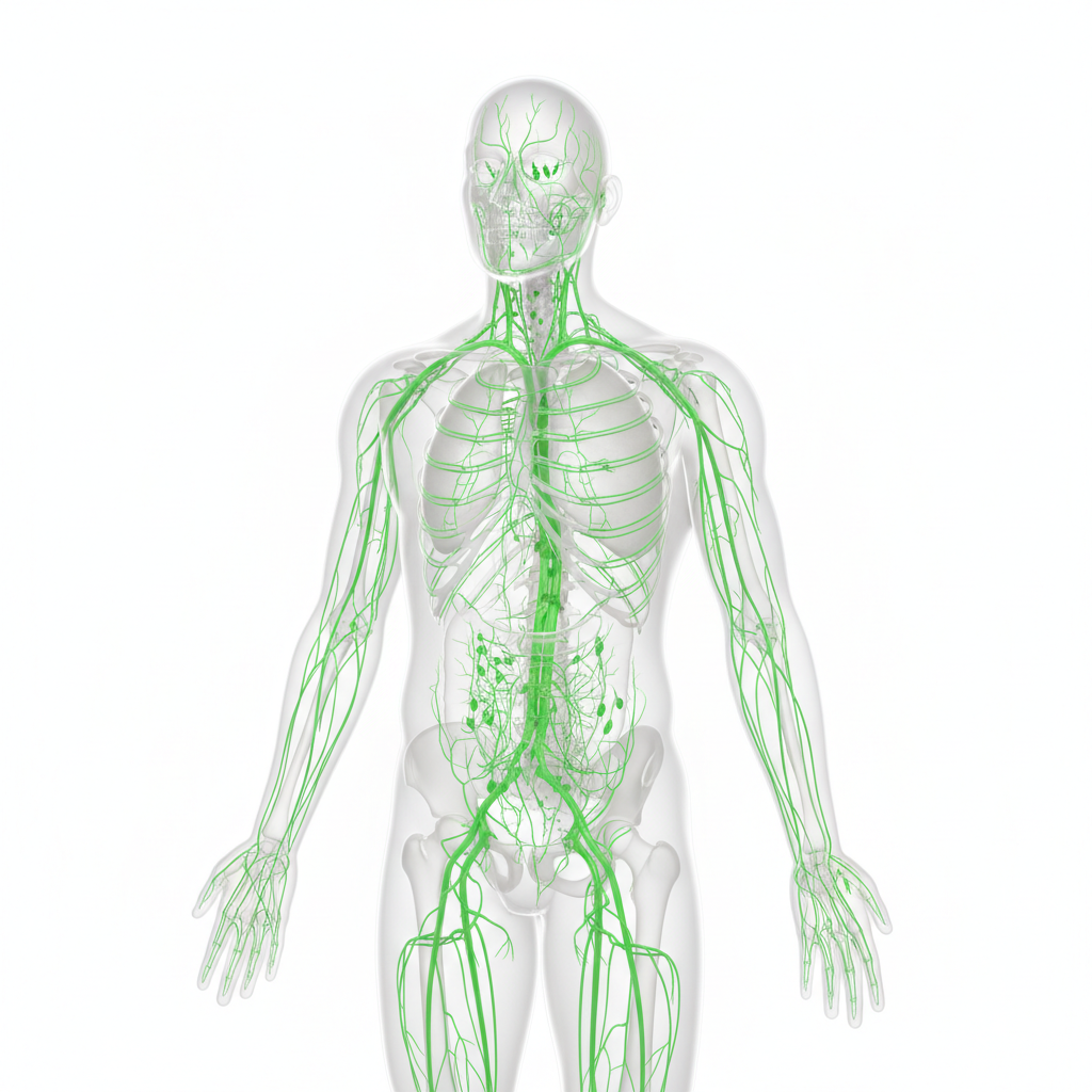
Lymphatic Capillaries
Microscopic, blind-ended tubes interweaving with blood capillaries.
Wall structure: Endothelial cells overlap to form one-way minivalves.
Anchoring filaments prevent collapse when tissue pressure rises.
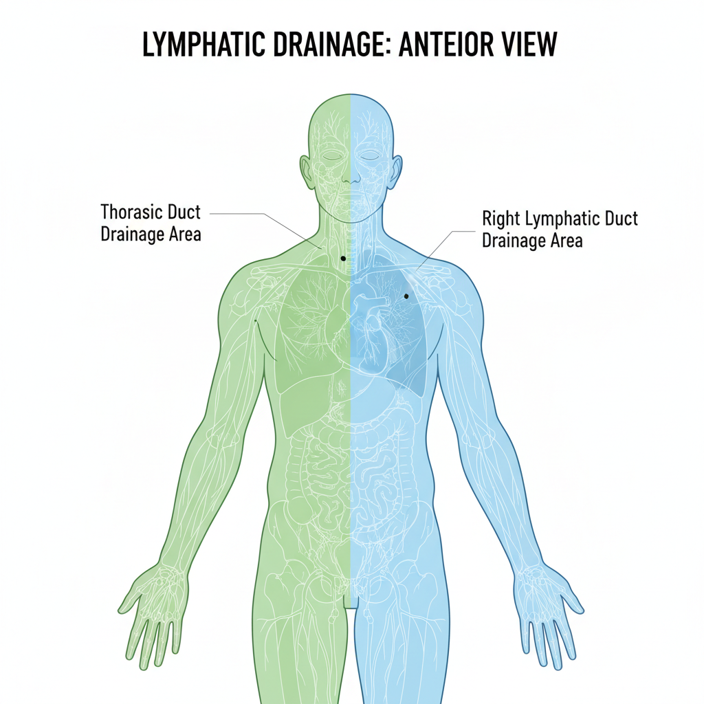
Lymphatic Trunks & Ducts
Right Lymphatic Duct: Drains the right upper arm and the right side of the head and thorax.
Thoracic Duct: Much larger; drains lymph from the rest of the body (legs, abdomen, left side of upper body).
Lymph Transport Mechanisms
The lymphatic system lacks a pump like the heart. Transport relies on:
Skeletal Muscle Pump: Milking action of active muscles.
Respiratory Pump: Pressure changes in thorax breathing.
Valves: Prevent backflow (similar to veins).
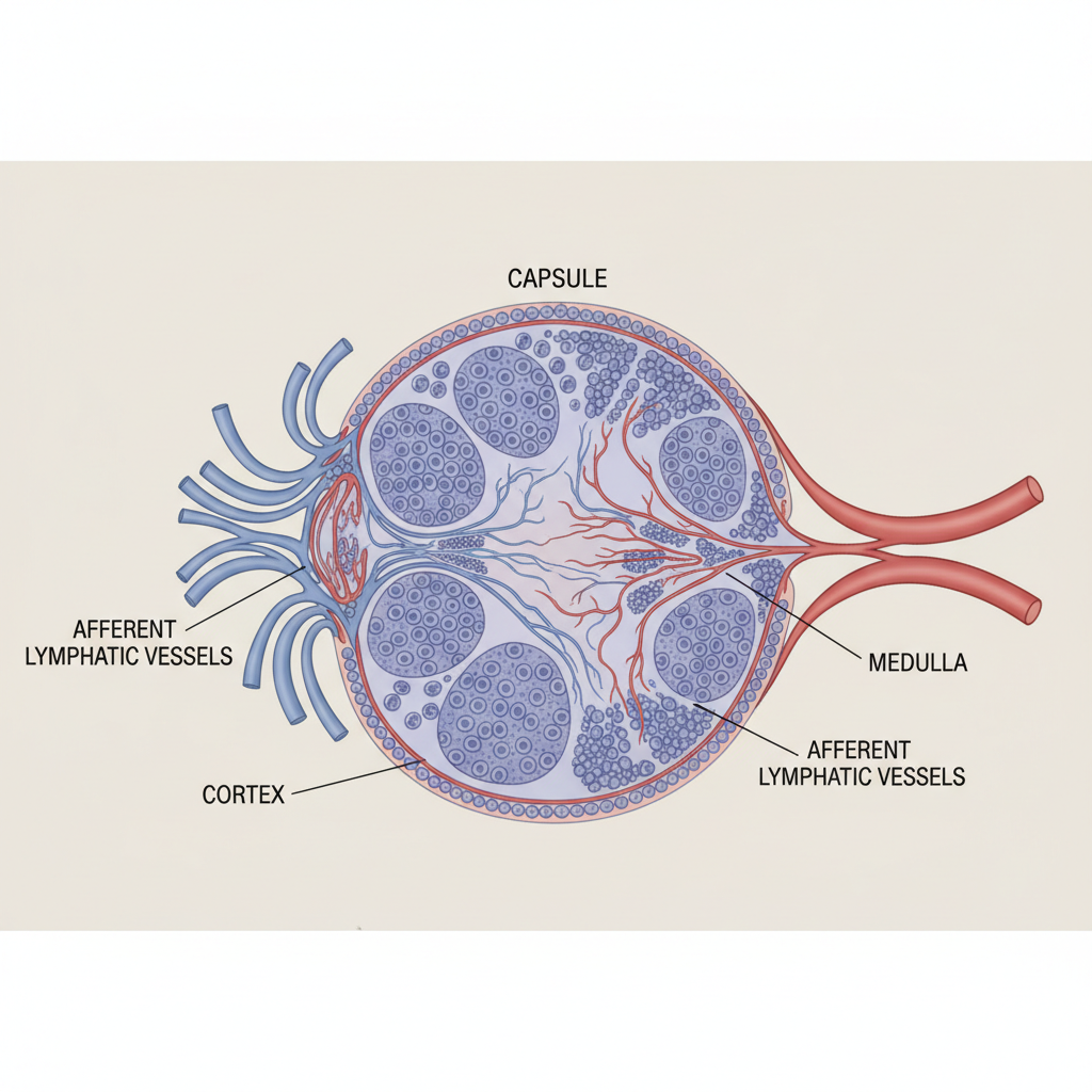
Lymph Node Anatomy
Bean-shaped filters clustered along lymphatic vessels.
Cortex: Contains follicles with Germinal Centers (B cell proliferation).
Medulla: Inner cords containing B cells and plasma cells.
Flow: Afferent vessels (in) > Sinuses > Efferent vessels (out).
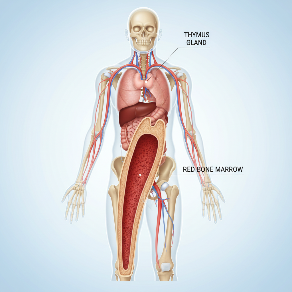
Primary Lymphoid Organs
Red Bone Marrow
Site where both B and T lymphocyte precursors originate. B cells mature here.
The Thymus
Located in the mediastinum. Site where T cells mature and become immunocompetent. Atrophies with age.
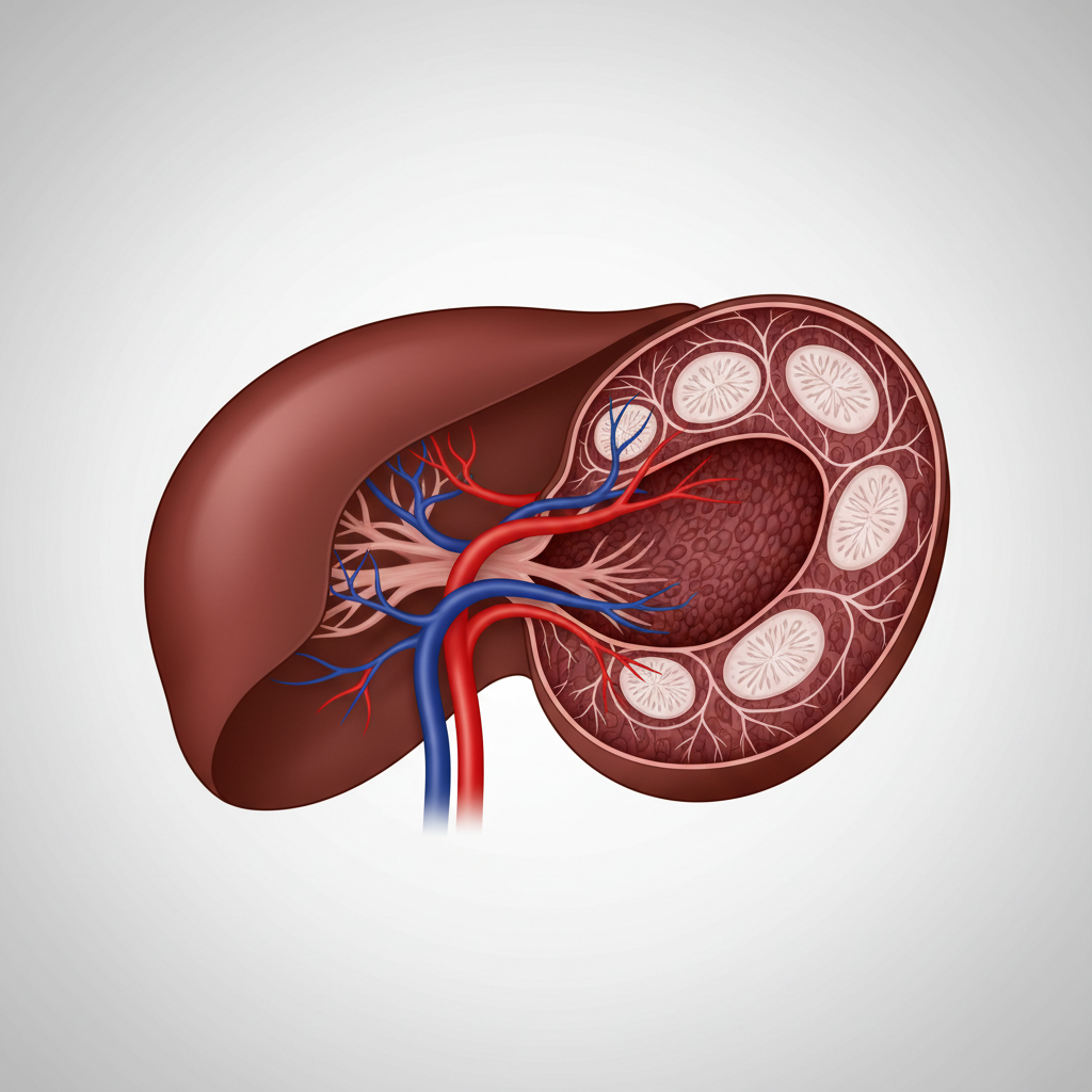
The Spleen
Largest Lymphoid Organ
Located in left upper quadrant of abdomen, curved around the stomach.
White Pulp: Immune function (lymphocytes on reticular fibers).
Red Pulp: Worn-out RBC destruction and blood storage.
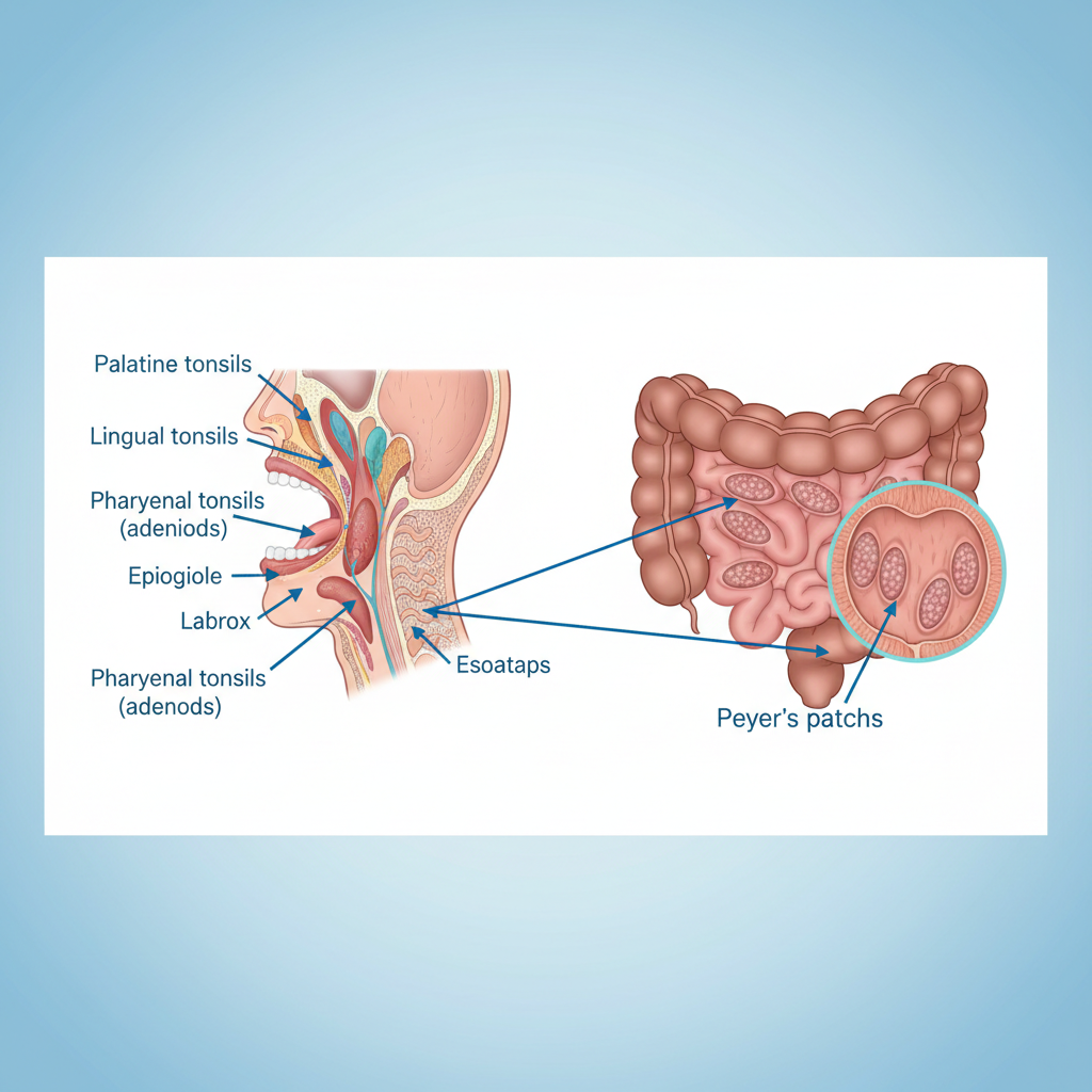
MALT & Tonsils
Mucosa-Associated Lymphoid Tissue
Protects mucous membranes in digestive, respiratory, and urinary tracts from foreign matter.
Tonsils: Ring of lymphoid tissue around pharynx (Palatine, Lingual, Pharyngeal).
Peyer's Patches: Clusters of lymphoid follicles in distal small intestine.
Summary: Vital Connections
The Lymphatic System is structurally connected to the Cardiovascular System (fluid return) and functionally connected to the Immune System (defense).
Failure of transport results in Edema (swelling).
- lymphatic-system
- human-anatomy
- physiology
- immunity
- lymph-nodes
- medical-education
- biology